
 Tap to zoom
What Is a Renal Cortical Cyst? Symptoms, Diagnosis, and Treatment
Renal cortical cyst | Simple vs. complex kidney cysts | Warning symptoms | Diagnosis and treatment | Needle drainage, laparoscopy, medication, and ablation
- Published on
- June 26, 2026
- Reading time
- 5 min read
- Last updated
- Updated: June 27, 2026
A renal cortical cyst is a fluid-filled cyst that develops in the outer part of the kidney, called the cortex. These cysts are usually found incidentally on ultrasound or CT scan.
Most renal cortical cysts are simple and benign. They rarely become cancerous or cause specific symptoms. Some people live comfortably for years without realizing they have a renal cortical cyst.
Urologic surgeon Kidney stone and urinary tract specialist
Note: To improve care quality and patient satisfaction, appointments are triaged by reason for visit. Each physician sees patients within the relevant urology subspecialty.
Book appointment
In this article, we explain renal cortical cysts, their symptoms, and their possible risks. We also review the methods used to diagnose and treat them. Read on for a clear overview of this type of kidney cyst.
What is a renal cortical cyst?
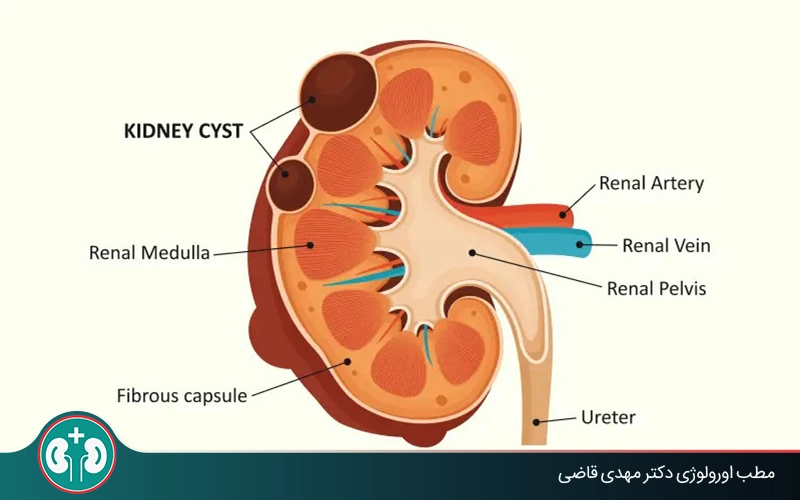
A renal cortical cyst is a small, round, closed sac filled with clear fluid or water that forms in the outer part, or cortex, of the kidney. To understand the definition of a renal cortical cyst more clearly, it helps to know that the kidney has two main parts:
The renal cortex is the outer layer of the kidney and contains millions of tiny filtering units called nephrons. This part is mainly responsible for cleaning the blood, removing waste products, and regulating the body’s water and electrolytes. Cysts that form in this area are renal cortical cysts.
The medulla is the inner part of the kidney. It is made up of tubules and renal pyramids, and it collects urine produced in the cortex and directs it toward the renal pelvis.
In simple terms, the cortex is the outer, active layer of the kidney, while the medulla is the inner part and part of the pathway that carries waste out. Knowing this difference helps explain why a renal cortical cyst specifically means a cyst on the surface portion of the kidney, not deep inside it or in another area.
Appointments related to Urologic surgeon Kidney stone and urinary tract specialist
Open the public booking path to review times and related information.
Book appointment
Simple renal cortical cyst vs. kidney cyst: what is the difference?
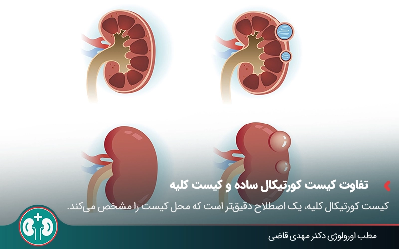
Kidney cyst is a general term for any cyst that forms anywhere in the kidney, whether in the cortex or the medulla.
Renal cortical cyst is a more specific term because it identifies where the cyst forms. These cysts arise only in the renal cortex, the outer and peripheral part of the kidney, which is why they are called cortical. Most cysts found incidentally on imaging are simple renal cortical cysts, which are among the most benign types of kidney cysts.
Simply put, every renal cortical cyst is a kidney cyst, but not every kidney cyst is cortical, because some may form in the medulla of the kidney.
Q&A — Urologic surgeon Kidney stone and urinary tract specialist
General questions are shown on the destination page after review.
Book appointment
Types of renal cortical cysts based on the Bosniak system
Most renal cortical cysts are simple, but accurate diagnosis is very important. Radiologists and doctors use a standard system called the Bosniak system to assess kidney cysts.
This system classifies cysts into categories based on their imaging features, such as wall thickness, septa, calcification, or solid material, to estimate the risk of malignancy or cancer. The table below reviews types of renal cortical cysts according to the Bosniak system.
Keep in mind that septa are thin walls, partitions, or dividers that form inside the fluid-filled space of a cyst. These walls divide the cyst into smaller sections or chambers.
Table: types of renal cortical cysts based on the Bosniak system | |||
|---|---|---|---|
Cyst type | Main features | Risk of malignancy | Need for follow-up/treatment |
Bosniak I (simple) | Thin, smooth wall; no septa or solid material | About 0% or benign | Follow-up and treatment are usually not needed. |
Bosniak II | May have very thin septa or small calcifications | Very low risk (less than 3%) | Periodic imaging with ultrasound or CT scan may be used, but long-term follow-up is usually not needed |
Bosniak IIF | Slightly thicker septa or several septa | Low risk (less than 5%) | Regular follow-up with periodic imaging |
Bosniak III | Thick walls, irregular septa, and suspicious solid components | Moderate risk (about 50%) | Often requires surgery, biopsy, or more detailed evaluation |
Bosniak IV | Clear solid components that contain blood vessels | High risk (more than 80%) | Requires surgery (removal of the mass or kidney) |
Symptoms and signs of a renal cortical cyst

In most cases, a renal cortical cyst causes no symptoms, and the person does not notice it at all. Many people are unaware of these cysts for years, and sometimes for life. Cortical cysts are often discovered incidentally during ultrasound or CT scans done for another medical reason.
Symptoms of large or multiple cortical cysts
When a cyst becomes large or when there are many cysts, symptoms may develop. These symptoms vary depending on the cyst’s location and how much pressure it puts on kidney tissue. The most important symptoms of large or multiple cysts include:
Flank or back pain: This pain is usually felt in the lower back, below the ribs, and on one side of the body. It can occur when the cyst becomes large enough to stretch the outer covering of the kidney or press on nearby tissues.
A feeling of fullness or heaviness in the abdomen: A large cyst can take up space in the abdomen and cause an uncomfortable feeling of fullness or heaviness.
Blood in the urine, or hematuria: Hematuria is uncommon and often happens because a cyst ruptures or bleeds. When hematuria occurs, the urine may look pink or red.
High blood pressure: Larger cysts, or cysts in certain locations, may press on kidney blood vessels and affect the mechanisms that regulate blood pressure. As a result, blood pressure may rise.
Infection: Sometimes a cyst can become infected. In that case, it may be associated with fever, severe pain, and tenderness over the kidney area.
Rare possible complications
In very rare cases, if a simple cortical cyst becomes very large or develops internal bleeding, it may lead to the following complications:
Bleeding inside the cyst;
Pressure on the kidney in some simple cysts larger than 10 cm.
Is a renal cortical cyst dangerous?

Most renal cortical cysts are benign and do not pose a danger to the person. These cysts usually grow slowly, but they are not cancerous and do not affect kidney function.
Only in a very small percentage of cases may a cyst be complex or show imaging features that raise concern for malignancy or kidney cancer. In these situations, the doctor uses CT scan or MRI and the Bosniak classification system to determine the cyst type.
The difference between left and right renal cortical cysts
Right-sided and left-sided cortical cysts do not differ in their nature or risk because of side alone. The only difference may be where pain or pressure is felt. For example, if the cyst is in the right kidney, the person may feel pain in the right flank. The nature and risk of the cyst depend only on its features, such as whether it is simple or complex.
In general, if an ultrasound or CT scan reports a cortical cyst, there is usually no need to worry. However, if you have symptoms such as persistent flank pain, fever, blood in the urine, or high blood pressure, it is best to see a urologist for a more detailed evaluation.
Methods for diagnosing a renal cortical cyst
Ultrasound, CT scan, and MRI are commonly used to diagnose a renal cortical cyst. Below is an overview of each method:
1. Ultrasound
Ultrasound is the first and most common method for diagnosing a cortical cyst. It is painless and noninvasive, and it can usually distinguish simple benign cysts from other kidney lesions. On ultrasound, a cyst appears as a round or oval cavity with a thin wall and clear fluid content. Ultrasound can also help the specialist identify the cyst’s exact location.
2. CT scan
If the ultrasound result is unclear, or if the cyst looks complex or suspicious for malignancy, the doctor may recommend a contrast-enhanced CT scan. This test helps evaluate the cyst’s internal features, wall thickness, calcification, or any solid mass. Based on these findings, the doctor classifies the cyst according to the Bosniak system.
3. MRI
In some cases, especially when CT scan cannot be performed, such as in patients who are sensitive to contrast material or in pregnant women, MRI is used. This method is highly accurate in distinguishing simple cysts from complex cysts and can show subtle changes in kidney tissue.

Treatment of a renal cortical cyst
In most cases, a renal cortical cyst does not need treatment, and periodic follow-up is enough. The specialist’s decision about treatment depends on the cyst’s size and location, whether symptoms are present, and whether there is any concern for malignancy. The main treatment approaches are described below.
1. Conservative treatment or active follow-up without surgery
If the cyst is small, simple, and benign, the best approach is conservative treatment or active follow-up. In this situation, the doctor monitors the cyst with periodic checkups, such as ultrasound every 6 to 12 months. If the cyst does not change in size or structure, no intervention is needed.
2. Medication treatment
There is no direct medication that makes the cyst disappear, but the doctor may prescribe medicines to control blood pressure or treat infection in the cyst. If the cyst becomes infected, antibiotics are used.
3. Needle drainage of the cyst
If the cyst is large or causes pain and pressure, minimally invasive treatments may be used. One minimally invasive option is needle drainage of the cyst, performed under ultrasound or CT guidance.
After drainage, a sclerosing agent, such as alcohol or a similar substance, is sometimes injected to help prevent the cyst from filling again. This substance damages the lining cells of the cyst wall and helps prevent new fluid secretion and re-enlargement of the cyst.
4. Laparoscopic surgery
If the cyst is larger, more complex, or recurrent, it may be removed from the surface of the kidney using minimally invasive laparoscopic surgery. This method is precise and usually has a shorter recovery period.
5. Ablation
In recent years, newer and less invasive treatments, such as radiofrequency ablation or laser-energy treatment, have been used to destroy selected cysts. Laser use for simple cysts is not very common. In general, these methods are usually considered when:
The patient is not a suitable candidate for laparoscopic surgery;
The cyst is in a special or deep location that makes surgical access difficult;
The cyst has caused significant pain or functional impairment, and simpler methods, such as needle drainage, have not been effective.
Summary
A renal cortical cyst is usually a benign lesion that forms in the outer part of the kidney, the cortex. In most cases, these cysts cause no symptoms, do not endanger a person’s health, and are found only during imaging checkups such as ultrasound. However, if they become large or multiple, they may cause flank pain, high blood pressure, or impaired kidney function.
A cortical cyst is diagnosed with ultrasound and, when needed, CT scan for accurate Bosniak classification. For simple cysts, the best approach is regular follow-up with a urologist and active periodic imaging to make sure no change occurs. If symptoms develop or malignancy is suspected, the doctor may recommend more specific treatments such as drainage, surgery, or minimally invasive therapy.
If you notice symptoms that may be related to a renal cortical cyst, you can see a urologist to identify the cause of the symptoms and receive appropriate treatment.
Frequently asked questions
Actions & related links
Related articles
All articlesWhat Is Polycystic Kidney Disease? (Fetal and Adult PKD)
What is polycystic kidney disease? Learn about inherited ADPKD and ARPKD, fetal and adult symptoms, complications, diagnosis, medicines, surgery, diet, fluids, and prevention-focused care.
Kidney Transplant: Cost, Blood Type Compatibility, and Surgical Method
What is kidney transplant? Learn about cost considerations, operation duration and method, diet, who may not be eligible, success rates, isolation precautions, and post-transplant care.
What Is Pyelonephritis? Kidney Infection Symptoms, Diagnosis, and Treatment
What is pyelonephritis? Learn kidney infection symptoms in women, children, men, and pregnancy; diagnosis, antibiotics, treatment, emergency warning signs, and cystitis differences.
Kidney Transplant in Children and Infants (Outcomes, Complications, Graft Life)
Kidney transplant in children and infants: age and weight readiness, dialysis versus transplant, growth and puberty, success rates, complications, graft life, medicines, vaccines, and follow-up care.
Comments
0 comments
No comments yet. Be the first to share your thoughts.