
 Tap to zoom
Prostate Ultrasound: Methods and Interpretation
How is prostate ultrasound performed? Types and methods, uses and importance, preparation, result interpretation, normal prostate volume, and findings that need follow-up.
- Published on
- June 26, 2026
- Reading time
- 5 min read
- Last updated
- Updated: June 27, 2026
Prostate ultrasound is an imaging method used to assess the size, shape, and condition of the prostate gland. Ultrasound is useful when current symptoms and earlier test results are not enough for a definite diagnosis, or when a biopsy is needed.
In this article, in simple but precise language, we explain how many types of prostate ultrasound there are and how each is performed. We also answer questions about what preparation prostate imaging needs, how results are interpreted, and which signs may require more serious follow-up.
Urologic surgeon Prostate disease specialist
Note: To improve care quality and patient satisfaction, appointments are triaged by reason for visit. Each physician sees patients within the relevant urology subspecialty.
Book appointment
If you have urinary symptoms or your doctor has ordered this test for you, this guide answers the questions you may have.
What Is Prostate Ultrasound?
Prostate ultrasound uses sound waves to create an image of the size and shape of the prostate and helps the doctor diagnose inflammation, benign prostatic enlargement, or cancerous masses.
Prostate ultrasound is usually performed for middle-aged or older men, especially when the patient has urinary symptoms or blood in the semen or when screening results are abnormal. In this situation, the urologist can use the ultrasound findings to make a more precise assessment.
Appointments related to Urologic surgeon Prostate disease specialist
Open the public booking path to review times and related information.
Book appointment
Prostate ultrasound is usually performed for the following purposes.
1. Biopsy to Evaluate the Possibility of Prostate Cancer
When the PSA level in the blood is high, the urologist feels a firm area on digital rectal examination of the prostate, and the MRI result is suspicious, a biopsy helps with diagnosing prostate cancer.
This is usually done under live transrectal ultrasound guidance, with fusion to MRI images. This approach increases biopsy accuracy and improves the chance of detecting small tumors. Ultrasound also helps the doctor see the position of ducts and tissue blood flow so the procedure or treatment can be carried out with minimal injury.
Q&A — Urologic surgeon Prostate disease specialist
General questions are shown on the destination page after review.
Book appointment

2. Diagnosing Benign Prostatic Hyperplasia (BPH)
In middle-aged and older men, ultrasound measures the gland volume accurately. If the prostate is enlarged, the doctor can also assess how much pressure it is placing on the urethra and decide whether medication is enough to manage benign prostatic hyperplasia or whether surgery is needed.
3. Identifying Inflammation or Infection (Prostatitis)
In cases where the patient has pelvic pain, burning with urination, or fever, ultrasound can show swelling, increased blood flow on Doppler, or tissue changes caused by inflammation.
4. Evaluating obstruction or abnormality of the urinary tract
Ultrasound helps the urologist determine whether the urine outlet is affected by obstruction, a cyst, calcification (calcium deposit), or abnormal protrusions. These findings can explain a feeling of incomplete emptying or frequent urination.
Types of Prostate Ultrasound and How They Differ
Different imaging methods are used to evaluate the prostate gland. The most important are transrectal ultrasound and abdominal prostate ultrasound. The main difference between these two methods is where the probe is placed and how accurate they are. Below, after a summary table, we introduce each method, its uses, advantages, and limitations.
Comparison Point | Transrectal Prostate Ultrasound | Abdominal Prostate Ultrasound |
|---|---|---|
How It Is Performed | Inserting a thin probe into the rectum | Placing the probe on the abdominal skin |
Accuracy | Acceptable; suitable for detailed evaluation and biopsy | Moderate; suitable for a general prostate evaluation |
Invasiveness | Minimally invasive; may be somewhat uncomfortable | Noninvasive and painless |
Preparation Needed | The bladder should be full (additional steps if biopsy is needed) | A full bladder |
Abdominal Prostate Ultrasound
In transabdominal prostate ultrasound, the ultrasound probe is placed with special gel on the skin of the lower abdomen. Sound waves pass through the abdominal wall toward the prostate area, and images of the prostate and bladder appear on the monitor.
This method is noninvasive and is performed in general imaging centers. Compared with transrectal prostate ultrasound, however, it is less accurate; image quality also depends partly on how full the bladder is.
The most important uses of this type of prostate ultrasound are:
Measuring overall prostate volume;
Checking residual urine in the bladder after urination;
Initial assessment of patients with mild urinary symptoms.

Transrectal Prostate Ultrasound (TRUS)
Transrectal prostate ultrasound (TRUS) is one of the methods used to evaluate the internal structure of the prostate and plays an important role in diagnosing prostate problems and performing biopsy. In this method, a thin covered probe with lubricating gel is inserted into the rectum to obtain close, detailed images of the prostate from behind.
This method can be combined with advanced techniques such as Doppler, elastography, and contrast; however, because the probe is inserted into the rectum, it may be somewhat uncomfortable for the patient.
This type of prostate ultrasound is used for:
Detailed evaluation of the internal structure of the prostate;
Identifying areas suspicious for cancer or inflammation;
Guiding the needle for diagnosis and biopsy;
Monitoring the treatment course of prostate-related diseases.
Preparing for Prostate Ultrasound
Conditions before prostate ultrasound may vary slightly depending on the type (abdominal or transrectal) and its purpose (for example, only checking prostate size or performing a biopsy).
Therefore, after your doctor orders the test and before you go to a radiology center, be sure to ask your doctor about the right timing for prostate ultrasound and the preparation you need.
Paying attention to the following points will help you be ready for prostate ultrasound:
Type of Ultrasound or Its Purpose | Point | Details |
|---|---|---|
Abdominal Prostate Ultrasound | Drinking water before the ultrasound | Better visualization of the prostate |
Transrectal prostate ultrasound, biopsy | Emptying the bowel with a laxative | Preventing infection |
Biopsy | Stopping blood-thinning medicines | Reducing the risk of bleeding |
Having a companion | If a sedative is used |
Steps in Prostate Ultrasound
The way prostate ultrasound is performed differs depending on whether it is abdominal or transrectal. Below, we explain how prostate ultrasound is done step by step in both methods:
Steps in Abdominal Prostate Ultrasound
Abdominal prostate ultrasound is a simple, quick, and painless method for evaluating the size, shape, and overall condition of the prostate. It usually takes about 20 to 30 minutes and is performed on the skin of the abdomen.
The steps in abdominal prostate ultrasound are as follows:
The radiologist asks you to wear comfortable clothing or a special ultrasound gown and lie down on the examination table.
A clear gel is applied to your lower abdomen so sound waves can transmit better.
The ultrasound probe is then placed on the abdomen and moved gently to obtain images from different angles.
The size, shape, and condition of the prostate are recorded in the images so the doctor can assess them.
If needed, the radiologist may ask you to empty your bladder and repeat the imaging.
After the examination, you wipe the gel from your skin and can return to your daily activities immediately.
Steps in Transrectal Prostate Ultrasound
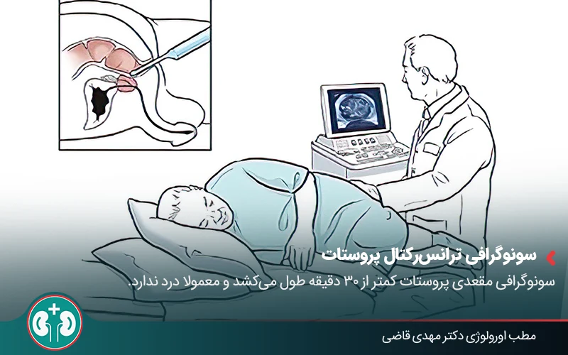
In transrectal ultrasound, a small probe is inserted into the rectum to obtain detailed images of the prostate.
This method usually takes less than 30 minutes, and if a biopsy is needed, it is performed during the same session.
The process is simple and generally tolerable, and proceeds as follows:
You put on a gown, lie on the examination table, and turn onto your side with your knees drawn toward your chest.
The technician prepares the special ultrasound probe with a sterile cover and lubricating gel.
The probe is gently inserted into the rectum until it reaches the area near the prostate.
Sound waves leave the probe and, after reflecting off prostate tissue, create its image on the monitor.
The radiologist moves the probe slightly to complete imaging from different angles.
If a biopsy is needed, the radiologist passes a thin needle through the probe into the prostate tissue and takes a small sample.
After imaging is finished, the radiologist removes the probe, and you can return to normal activities after a short rest.
Position of the second image
Does Prostate Ultrasound Hurt?
Prostate ultrasound is one of the lowest-risk methods for checking prostate health and is usually not painful. It is performed without surgery, without radiation, and without general anesthesia.
Only in transrectal ultrasound is a narrow, soft device inserted into the rectum to image the prostate. This may cause a feeling of pressure or mild discomfort, but it is usually not painful, and many people describe the sensation as similar to a simple rectal exam.
If the doctor decides to take a prostate sample during the ultrasound (biopsy), a thin needle may be used to obtain the sample. In this situation, even with local anesthesia, you may still feel mild pain or discomfort.
Side Effects of Prostate Ultrasound
Abdominal ultrasound is noninvasive and has no side effects. Transrectal prostate ultrasound (TRUS) by itself is also usually safe and may cause only mild rectal discomfort or a feeling of pressure. If transrectal ultrasound is combined with prostate biopsy, temporary bleeding, blood in the urine or stool, or mild infection may occur. As noted, however, these are complications of prostate biopsy, and transrectal prostate ultrasound alone does not cause them.
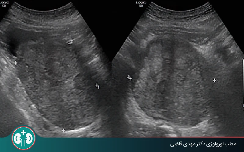
Interpreting Prostate Ultrasound Results and Follow-Up
After ultrasound is performed, the report is usually interpreted by a radiologist and given to the treating doctor.
For many patients, however, the terms and numbers in the report can be confusing. Here are the key points to look for in a prostate ultrasound report.
Normal Prostate Size on Ultrasound

The normal prostate size in young men is usually about 20 to 30 milliliters, and this volume gradually increases with age. Put more simply, normal prostate dimensions on ultrasound are roughly 3 x 3 x 5 cm. If the prostate volume is reported as higher than this, the ultrasound report may use the term enlarged prostate.
In many cases, this does not mean cancer or a dangerous disease. The most common cause of prostate enlargement is a condition called benign prostatic hyperplasia (BPH). On prostate ultrasound, BPH is usually seen in men older than 50 to 60 years and is part of the natural aging process.
In the following situations, the doctor orders additional tests for further evaluation:
If the prostate volume is significantly higher than the normal prostate size on ultrasound.
If the patient has symptoms such as frequent urination, burning with urination, or a weak urine stream.
Measuring post-void residual urine, or PVR, is one of these follow-up evaluations. This type of ultrasound measures the amount of urine left in the bladder after it has been completely emptied, in order to assess urinary system function in a person with prostate problems.
Suspicious Signs in the Ultrasound Report
Some terms seen in prostate ultrasound reports may point to inflammation, abnormal enlargement, or even a cancerous mass.
Below are common prostate ultrasound report terms:
1. Hypoechoic Lesion
A hypoechoic lesion is one of the common findings that may raise concern for prostate cancer on prostate ultrasound. This area appears darker than the surrounding tissue. However, you should know that:
1. Many cancers are not visible on ultrasound, so ultrasound alone is not enough for a definite answer.
2. Some hypoechoic lesions are benign; therefore, do not worry just because you see this term in the ultrasound report, and leave the final diagnosis to the urologist.
2. Enlarged Prostate
In prostate ultrasound, enlarged means that the prostate volume is greater than normal. This condition can occur because of benign enlargement (BPH) or other causes.

3. Heterogeneous Tissue
Heterogeneous tissue means the prostate tissue is not uniform and even, and it may have inflammatory, fibrotic, or even cancerous changes.
4. Irregular Margins
Irregular margins mean that the prostate has borders that are not smooth or even, which sometimes accompanies abnormal or invasive growth; however, by itself it is not a reliable criterion for cancer.
5. Increased Blood Flow
A report of increased blood flow is a finding seen on Doppler ultrasound of the prostate and may indicate inflammation, infection, or an active mass.
6. Protrusion
A protrusion or bulge in the wall or a specific part of the prostate may indicate abnormal tissue growth. In some cases, this can be due to a compressive mass or a lesion that changes the normal structure of the gland.
7. Calcification
Bright or white spots on prostate ultrasound indicate calcium deposits in the tissue. These deposits usually result from chronic inflammation or an old infection, but in some cases they can be more important when seen alongside other findings.
8. Prostate Cyst
On ultrasound, a cyst appears as a dark, fluid-filled area. This is usually benign and may occur because of duct blockage or congenital changes, but if it is large or pressing on the urethra, it needs more careful evaluation.
Ultrasound alone is not a reliable method for diagnosing prostate cancer. It is used more often for purposes such as measuring prostate volume, guiding biopsy, evaluating urinary retention, and assessing the anatomy of the gland.
Are Additional Tests or Procedures Needed?
Yes. In many cases, prostate ultrasound is only one tool for evaluating this gland, and the doctor may also use other tests or diagnostic methods to complete the assessment. The choice of these evaluations depends on why the ultrasound was performed, the patient's symptoms, and the results obtained from tests.
One common test in this area is measuring prostate-specific antigen (PSA) in the blood. A higher PSA level can be seen in several conditions, such as benign prostatic hyperplasia (BPH), prostate inflammation (prostatitis), or prostate cancer. Therefore, this test helps the doctor perform more detailed evaluation if needed.
If test results or imaging findings need further evaluation, the doctor may use the following methods:
Transrectal ultrasound-assisted biopsy (TRUS-guided biopsy): precise sampling from suspicious parts of the prostate to check for abnormal cells.
MRI: to see the internal tissues of the prostate more clearly and better identify the location and extent of a lesion.
Repeat ultrasound or PSA testing: in some cases, periodic evaluations are performed to assess the pattern of change.
Overall, doctors usually consider digital rectal examination of the prostate, ultrasound results, PSA level, MRI findings, and biopsy results if needed to gain a more complete picture of prostate status and, based on that, choose the best treatment or follow-up plan for the patient.
Summary
Prostate ultrasound is one of the common and reliable methods for evaluating this gland in men. It helps the doctor assess its size, structure, and possible changes. This evaluation is usually performed by two main methods: abdominal prostate ultrasound and transrectal prostate ultrasound (TRUS), each of which has different uses and levels of accuracy.
Abdominal ultrasound is used more often for general evaluation of prostate size and assessment of residual urine in the bladder.
Transrectal ultrasound provides more detailed images of prostate tissue and is useful in situations such as evaluating suspicious areas or guiding biopsy.
Finally, the results of this imaging are usually reviewed alongside tests such as PSA, the clinical examination, and MRI when needed, so the urologist can make a more accurate diagnosis of problems such as benign prostatic hyperplasia, inflammation, or possible lesions, and choose the best treatment or follow-up path for the patient.
Frequently Asked Questions
References
Actions & related links
Related articles
All articlesWhat Is Polycystic Kidney Disease? (Fetal and Adult PKD)
What is polycystic kidney disease? Learn about inherited ADPKD and ARPKD, fetal and adult symptoms, complications, diagnosis, medicines, surgery, diet, fluids, and prevention-focused care.
What Is a Renal Cortical Cyst? Symptoms, Diagnosis, and Treatment
Renal cortical cyst | Simple vs. complex kidney cysts | Warning symptoms | Diagnosis and treatment | Needle drainage, laparoscopy, medication, and ablation
Kidney Transplant: Cost, Blood Type Compatibility, and Surgical Method
What is kidney transplant? Learn about cost considerations, operation duration and method, diet, who may not be eligible, success rates, isolation precautions, and post-transplant care.
What Is Pyelonephritis? Kidney Infection Symptoms, Diagnosis, and Treatment
What is pyelonephritis? Learn kidney infection symptoms in women, children, men, and pregnancy; diagnosis, antibiotics, treatment, emergency warning signs, and cystitis differences.
Comments
0 comments
No comments yet. Be the first to share your thoughts.