
 Tap to zoom
Prostate Cancer Diagnosis: From PSA Blood Test to Biopsy
Prostate cancer diagnosis | PSA test, DRE, urine tests, ultrasound, MRI and biopsy | Early diagnosis, steps, accuracy and next tests | Dr. Mahdi Ghazi Clinic
- Published on
- June 26, 2026
- Reading time
- 5 min read
- Last updated
- Updated: June 27, 2026
Diagnosis of prostate cancer as early as possible greatly improves the chance of successful treatment. It is one of the most common cancers in men, and the important point is that it usually causes no clear symptoms in the early stages. For this reason, if you develop urinary symptoms, you should see a doctor.
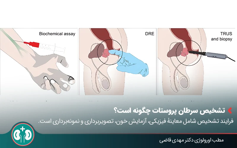
Diagnosing prostate cancer usually requires several methods. After a PSA blood test and a digital rectal examination, if the results are suspicious, the doctor uses other tests to make a definite diagnosis. With ultrasound, prostate MRI, and finally tissue sampling (biopsy), the doctor can reach a final and accurate diagnosis.
Urologic surgeon Prostate disease specialist
Note: To improve care quality and patient satisfaction, appointments are triaged by reason for visit. Each physician sees patients within the relevant urology subspecialty.
Book appointment
In this article, we review the pathway for diagnosing prostate cancer step by step: what role each method plays, when it is recommended, what its benefits and limitations are, and who is considered high risk.
If you want to know when a raised PSA is concerning, how MRI helps with decision-making, and when a biopsy is truly needed, continue reading.
How Is Prostate Cancer Diagnosed?
In general, the diagnostic process for prostate cancer is as follows:
Appointments related to Urologic surgeon Prostate disease specialist
Open the public booking path to review times and related information.
Book appointment
Review of symptoms and clinical examination: assessment of urinary problems and possible changes on prostate examination;
Blood test: measurement of PSA (prostate-specific antigen);
Imaging: closer evaluation of the prostate with MRI or transrectal ultrasound,
Tissue sampling (biopsy): the definitive method for confirming or ruling out cancer and determining the risk level of the disease.
Below, we will review all diagnostic methods and tests for this disease in more detail.
Digital Rectal Examination (DRE)
Digital Rectal Examination (DRE) is one of the older and simpler ways to assess prostate health. By feeling the prostate through the rectum, the urologist can evaluate the following:
Prostate size: whether the gland is normal-sized or enlarged;
Prostate consistency: whether the tissue feels soft, firm, or uneven;
Prostate surface: whether it is smooth or has raised and irregular areas;
A lump or abnormal hardness: which may be a warning sign.
This examination is quick, requires no special preparation, and is usually painless. Keep in mind that DRE alone is not enough to definitively diagnose cancer, but it can help decide whether further testing is needed.
Q&A — Urologic surgeon Prostate disease specialist
General questions are shown on the destination page after review.
Book appointment
What Is a PSA Blood Test and What Is It Used For?

The PSA test is one of the first and most accessible tools for assessing the possibility of prostate cancer. It measures the level of prostate-specific antigen in the blood, a protein naturally produced by prostate cells.
In general, a PSA value below 4 nanograms per milliliter is often considered within the expected range, although this number can vary with age and a person's overall health. An elevated PSA raises the possibility of a prostate problem and usually prompts the doctor to consider more detailed evaluation, such as imaging or, in some cases, biopsy.
However, a high PSA by itself does not mean cancer, because conditions such as inflammation or benign prostatic hyperplasia can also raise PSA. For this reason, the PSA test is used as a screening tool, and its result should always be interpreted along with other clinical and diagnostic findings.
Urine Testing in Prostate Cancer Diagnosis
Diagnosing prostate cancer with a urine test is usually done as an additional test. In this test, urine is checked for biomarkers or specific genes. Some of these genes are more active in prostate cancer cells and can indicate a higher likelihood of cancer.
This method can help the doctor decide whether a biopsy is needed. Its main advantage is that it is noninvasive and may help avoid an unnecessary costly or uncomfortable test.
Imaging Methods in Prostate Cancer Diagnosis
Physical examination and blood tests are not always enough to diagnose prostate cancer. Doctors also use imaging methods to identify the exact location of a tumor and the extent of disease progression.
The most important of these methods are prostate ultrasound, prostate MRI, CT scan, and nuclear medicine scans. Each has its own benefits and limitations, and the doctor chooses among them based on the patient's condition.
Transrectal Ultrasound (TRUS)
Diagnosing prostate cancer with transrectal ultrasound (TRUS) is one of the older and commonly used methods. In this procedure, a small probe is inserted through the rectum to give the doctor a clear image of the prostate.
If the doctor sees a suspicious area on ultrasound, a biopsy can be done at the same time for further evaluation.
Benefits of transrectal ultrasound:
Does not require surgery and is generally safe;
Quick and relatively easy to perform;
Can be combined with biopsy during the same session;
Shows prostate volume and size.
Limitations of transrectal ultrasound:
It does not show all tumors clearly;
Some benign and cancerous changes can look similar.
In simple terms, transrectal ultrasound is mostly an initial tool: it helps determine prostate size and general changes or makes biopsy more precise. However, compared with prostate MRI, ultrasound provides less detail and lower imaging accuracy.
The Role of MRI in Prostate Cancer Diagnosis
Today, prostate MRI is regarded as the most accurate imaging method for local evaluation of the prostate. It can show the exact location of a tumor, its size, and even whether cancer has extended beyond the prostate.
Preparation before MRI is important. To help MRI (magnetic resonance imaging) produce accurate and clear images, it is best to follow a few simple instructions before the test:
Emptying the bowel before MRI to reduce shadowing and extra movement;
Avoiding a heavy meal for several hours before imaging;
In some cases, taking an antispasmodic medication as directed by the doctor to keep the prostate and bowel more still,
Removing metal objects, such as a belt, watch, or jewelry, before entering the MRI scanner.
Benefits of MRI in Prostate Cancer Diagnosis
Some of the most important benefits of MRI in prostate cancer diagnosis include:
High image resolution and detection of small or hidden lesions;
Assessment of whether the tumor has spread outside the prostate;
Helping choose the most appropriate treatment strategy, such as surgery, radiation therapy, or active surveillance;
Monitoring changes and growth of the tumor in low-risk cancers;
Improving biopsy accuracy by combining MRI images with live ultrasound images (MRI-ultrasound fusion prostate biopsy)

CT Scan and Nuclear Medicine Scan
When the doctor needs to know whether prostate cancer has spread to other parts of the body, these two methods may be used:
1. CT scan: A CT scan is not very useful by itself for diagnosing the prostate tumor, because small tumors are not shown well. However, when the goal is to check whether cancer has spread to lymph nodes or other organs, CT can be useful.
2. Nuclear medicine scan: A nuclear medicine scan is used to detect spread of cancer to the bones or other parts of the body. Newer methods use radiotracers, substances that attach to cancer cells and show the doctor exactly where the cancer has spread on the images.

Prostate Biopsy and Interpretation of Its Results
When the PSA blood test or the doctor's digital rectal examination shows a suspicious finding, prostate biopsy is used for a definitive diagnosis.
In addition to diagnosing whether cancer is present, prostate tissue sampling helps determine the severity of the disease and how fast it may be progressing. Next, we review how biopsy is performed and the different biopsy methods.
How Is a Prostate Biopsy Performed?
In general, after local anesthesia, the doctor uses thin needles to remove several small samples of prostate tissue. These samples are examined under a microscope to determine whether the cells are normal or cancerous.
The biopsy procedure itself usually takes 15 to 20 minutes, and during it the doctor takes about 10 to 12 small samples.
As with any medical procedure, biopsy can have side effects, including blood in the urine or semen, mild pain, or a small risk of infection; these are often mild. Below, we review the different methods.
Prostate Biopsy Methods
Transrectal biopsy: The most common route for sampling the prostate is transrectal biopsy, which is performed by inserting a small probe into the rectum. In this method, the doctor uses transrectal ultrasound imaging to guide the needle into the prostate and take tissue samples.
Transperineal biopsy: Another method of prostate sampling is transperineal biopsy. In this approach, the doctor passes the needle through the skin between the anus and the scrotum to reach the prostate and collect samples.

Gleason Score in Prostate Cancer Diagnosis
After sampling, the pathologist or laboratory doctor examines the tissue under a microscope. The biopsy result is usually reported using a measure called the Gleason score. As shown in the table below, this score indicates how normal or abnormal the cells look:
Gleason score | Meaning |
|---|---|
Score 6 (3+3) | Low-risk cancer; the cells look similar to normal cells. |
Score 7 (3+4) | Lower intermediate risk |
Score 7 (4+3) | Higher intermediate risk |
Score 8 to 10 | High-risk cancer; the cells look clearly abnormal and aggressive. |
As you saw in the table, biopsy results sometimes include two numbers written together, such as 3+4 or 4+3. This means the doctor has seen two main cell patterns. In this situation, if the first number is higher, the cancer is more aggressive.
In simple terms, interpreting biopsy results with the Gleason score helps the doctor decide whether the patient only needs monitoring or needs more active treatment, such as surgery or radiation therapy.
Suspicious Results and Next Steps
Sometimes biopsy results are suspicious, meaning the cells have changed but it is not yet clear whether they are cancerous.
Two common suspicious results in this situation are:
Abnormal but noncancerous changes in prostate cells (HGPIN): This means cancer has not yet developed, but the cells show signs of change and should be monitored regularly.
A group of suspicious cells in the prostate (ASAP): In this situation, the doctor sees that some cells are not normal and the chance of cancer is higher; for this reason, a repeat biopsy is usually done.
If the biopsy result is unclear, the doctor usually recommends the following additional tests:
Prostate MRI to evaluate suspicious areas more clearly;
Repeat biopsy with greater precision;
In some cases, newer blood or genetic tests.
What Is the Best Method for Diagnosing Prostate Cancer?
Prostate cancer diagnosis is a step-by-step process. Doctors usually use a combination of several methods to make sure the result is accurate and reliable.
The PSA blood test, digital rectal examination, imaging such as MRI, and finally biopsy each play a specific role in this pathway; however, the definitive result comes from biopsy.
Comparing these methods shows that, because each test has limitations, the best way to diagnose prostate cancer is usually a combination of tests, not a single method. Below is a table comparing the main prostate cancer diagnostic methods:
Method | Approximate accuracy | Benefits | Limitations |
|---|---|---|---|
PSA blood test | Moderate | Simple, accessible, noninvasive | False-positive and false-negative results; influenced by age and health conditions |
Digital rectal examination | Low | Quick; no special equipment needed | Detects only larger lumps or surface changes in the prostate |
Transrectal ultrasound | Moderate | Can be combined with biopsy; measures prostate volume | Low sensitivity for directly detecting cancer |
CT scan | Moderate | Evaluates lymph nodes and metastasis (spread of cancer to other areas) | Low accuracy for lesions inside the prostate |
Nuclear medicine scan | High | Best method for detecting spread of cancer to bone | Used only in high-risk patients and not useful for initial diagnosis |
Prostate MRI | High | Detailed image of tumor location and size | Higher cost; requires preparation |
Prostate biopsy | Definitive | The only method that confirms the presence of cancer | Invasive; possible mild complications such as bleeding |
Importance of Early Prostate Cancer Diagnosis
Prostate cancer is one of the most common cancers in men and usually has no specific symptoms in the early stages. For this reason, prostate cancer screening can save lives, because the earlier the disease is found, the more effective treatment can be.
Screening
With prostate cancer screening, the disease can be detected in its early stages. This is especially important for men with a family history or genetic changes:
A PSA blood test is usually started at age 50, and at age 45 or even 40 in high-risk men.
Physical prostate examination (DRE) is no longer considered an independent screening method, but alongside PSA it can help with earlier diagnosis.
Awareness of Prostate Cancer Risk Factors
Understanding prostate cancer risk factors helps the doctor decide more easily who should start screening earlier or undergo more detailed evaluation.
The most important factors include:
Older age, especially after age 50;
Family history of prostate cancer, such as an affected father or brother;
Inherited genetic changes, such as a BRCA2 gene mutation;
Unhealthy lifestyle factors, such as obesity, smoking, physical inactivity, and a high-fat diet.

Summary
Prostate cancer cannot be diagnosed reliably with a single test alone; a combination of key methods provides more dependable results.
A PSA blood test is the starting point for evaluation, and if abnormal values are found, the doctor usually uses imaging such as prostate MRI to identify the location and extent of a mass. Ultimately, prostate biopsy is the definitive way to confirm or rule out cancer, and the severity of the disease is also determined by examining these samples.
Because prostate cancer usually has no clear symptoms in the early stages, it is important to pay attention to warning signs such as urinary problems or blood in the urine.
In the end, seeing a doctor at the right time and completing the necessary evaluations can significantly increase the chance of successful treatment.
Frequently Asked Questions
Actions & related links
Related articles
All articlesWhat Is Polycystic Kidney Disease? (Fetal and Adult PKD)
What is polycystic kidney disease? Learn about inherited ADPKD and ARPKD, fetal and adult symptoms, complications, diagnosis, medicines, surgery, diet, fluids, and prevention-focused care.
What Is a Renal Cortical Cyst? Symptoms, Diagnosis, and Treatment
Renal cortical cyst | Simple vs. complex kidney cysts | Warning symptoms | Diagnosis and treatment | Needle drainage, laparoscopy, medication, and ablation
Kidney Transplant: Cost, Blood Type Compatibility, and Surgical Method
What is kidney transplant? Learn about cost considerations, operation duration and method, diet, who may not be eligible, success rates, isolation precautions, and post-transplant care.
What Is Pyelonephritis? Kidney Infection Symptoms, Diagnosis, and Treatment
What is pyelonephritis? Learn kidney infection symptoms in women, children, men, and pregnancy; diagnosis, antibiotics, treatment, emergency warning signs, and cystitis differences.