
 Tap to zoom
What Is Prostate Fusion Biopsy and When Is It Used?
Learn how MRI/ultrasound fusion prostate biopsy works, when it is recommended for suspected prostate cancer, possible risks, recovery, and cost.
- Published on
- June 26, 2026
- Reading time
- 5 min read
- Last updated
- Updated: June 27, 2026
Prostate fusion biopsy is a modern method for diagnosing prostate cancer that combines MRI and ultrasound imaging. Compared with traditional biopsy, this method offers greater accuracy, helps identify suspicious areas in the prostate, and can reduce the need for repeat biopsies.
In this article, we provide a comprehensive overview of prostate fusion biopsy, including its benefits and uses, how the procedure is performed, possible side effects, costs, and its role in the future of prostate cancer diagnosis. Read on to learn more about this method.
Urologic surgeon Prostate disease specialist
Note: To improve care quality and patient satisfaction, appointments are triaged by reason for visit. Each physician sees patients within the relevant urology subspecialty.
Book appointment
Introducing Prostate Fusion Biopsy
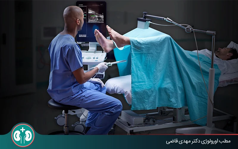
The newer prostate biopsy method, known as fusion biopsy, works by linking and overlaying detailed MRI images with ultrasound. First, the patient has a prostate MRI to identify areas suspicious for cancer. Then, during the biopsy, advanced software fuses the MRI images with live ultrasound images.
In practice, the MRI images are transferred to a special ultrasound system, and the urologist can see the MRI images live and simultaneously on the ultrasound screen while performing the biopsy.
This image overlay gives the urologist a precise three-dimensional map of the prostate. By seeing the exact location of suspicious areas, the biopsy needle can be guided to those areas with millimeter-level accuracy. This targeted approach significantly increases the chance of finding cancer cells and minimizes the need for broad random sampling.
Appointments related to Urologic surgeon Prostate disease specialist
Open the public booking path to review times and related information.
Book appointment
The precise, lower-error mechanism of prostate fusion biopsy distinguishes it from older methods and has made it one of the most reliable approaches for diagnosing prostate cancer.
The Role of MRI in Fusion Biopsy
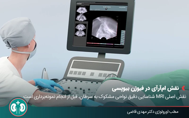
The accuracy of prostate MRI fusion is one of the most important factors in the success of this biopsy method. In fusion biopsy, MRI's main role is to identify areas suspicious for cancer before tissue sampling is performed.
Unlike ultrasound, which can show only general structural changes, multiparametric MRI can assess detailed features of prostate tissue, such as blood flow and cellular density. This information helps the radiologist identify and map areas with a higher probability of cancer.
Q&A — Urologic surgeon Prostate disease specialist
General questions are shown on the destination page after review.
Book appointment
This precise mapping greatly reduces diagnostic error and helps prevent false-negative biopsies, meaning failure to detect cancer when it is present.
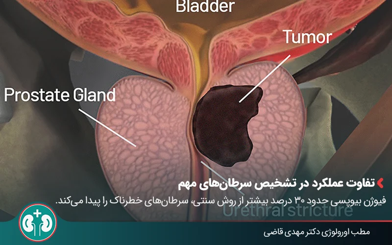
Difference Between Older Biopsy Methods and Prostate Fusion Biopsy
In the past, prostate biopsy was performed with the TRUS method under transrectal ultrasound guidance. In this approach, the doctor could see only the general size and shape of the prostate and, without a precise view of suspicious areas, would take several samples randomly from different parts of the prostate gland.
By contrast, prostate fusion biopsy combines detailed MRI and ultrasound images, enabling targeted sampling for the doctor.
Traditional Biopsy vs. Prostate Fusion Biopsy | ||
|---|---|---|
Criteria | Traditional TRUS Biopsy | Prostate Fusion Biopsy |
Accuracy | Lower, with a higher chance of error | High, with precise identification of suspicious areas |
Number of Samples | Higher (12-14 samples, depending on the urologist's expertise) | Fewer, targeted, and selective |
Chance of Error | High, with a risk of false negatives | Much lower because of precise targeting |
Cost | Lower in the short term | Higher initially, but more suitable over the long term |
Side Effects | More likely because more samples are taken | Less likely because repeat sampling is reduced |
Benefits of Prostate Fusion Biopsy Compared with Traditional Biopsy
Prostate fusion biopsy has several advantages over traditional tissue sampling. In this section, we outline some of the most important benefits of this modern method:
Greater accuracy: The most important benefit of this method is its excellent accuracy. By directly targeting suspicious areas identified on MRI, fusion biopsy can significantly increase the chance of detecting clinically significant cancers.
Targeted sampling: Instead of taking many untargeted samples, this newer method samples areas with the highest likelihood of containing cancer cells. This helps avoid unnecessary sampling of healthy tissue.
Reduced need for repeat biopsy: Because fusion biopsy is highly accurate, the likelihood of needing a repeat biopsy later because of a false-negative result is reduced. This can lower the patient's anxiety and stress and may also avoid repeated, additional costs and side effects.
Cost Comparison: Prostate Fusion Biopsy and TRUS
In terms of cost, the traditional method is usually less expensive than fusion biopsy; however, because its accuracy is lower, it may need to be repeated. Therefore, prostate fusion biopsy is more expensive at first, but with more accurate diagnosis and a reduced need for repeat biopsy, it may be more cost-effective over time.
Difference in Detecting Clinically Significant Cancers
The U.S. National Institutes of Health (NIH) began a study from 2007 to 2014 involving more than 1,000 men suspected of having prostate cancer. These men underwent both the traditional method (TRUS biopsy) and the newer MRI/ultrasound fusion method so the results could be compared.
The results showed that traditional TRUS detected only 122 cases of high-grade/high-risk prostate cancer, whereas fusion biopsy found 173 cases of the same high-grade/high-risk cancers. In other words, fusion biopsy detected about 30% more dangerous prostate cancers than the traditional method.
Who Needs Fusion Biopsy?
Fusion-based prostate cancer diagnosis is especially recommended for men who need a more precise assessment for prostate cancer. It is a strong option for people with suspicious laboratory or imaging findings, including:
Men with a high PSA (prostate-specific antigen) level: If the PSA level in the blood remains high or rises quickly, it may be a sign of prostate cancer. In this situation, the urologist may recommend fusion biopsy for a more detailed evaluation.
Suspicious findings on prostate MRI: If MRI shows suspicious areas in the prostate, fusion biopsy is an ideal way to precisely target and sample those areas.
Family history of prostate cancer: Men with a strong family history of prostate cancer may be at higher risk. In these men, even when PSA is relatively normal, fusion biopsy may help with early diagnosis if symptoms are present and the urologist considers it appropriate.
Previous unsuccessful biopsy: One of the most common reasons to recommend fusion biopsy is a negative result from previous older biopsies despite persistently high PSA or other signs. In this situation, the doctor may conclude that older sampling methods did not find hidden cancerous areas, and fusion biopsy, because of its higher accuracy, may help identify the disease.
How Fusion Biopsy Is Performed
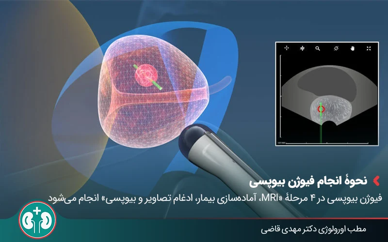
Fusion biopsy is performed in four stages: MRI imaging, patient preparation, image fusion, and tissue sampling. This process is performed with local anesthesia, although in some cases the doctor may decide that general anesthesia is needed. The stages of fusion biopsy are explained below:
MRI imaging: The patient first undergoes prostate MRI so areas suspicious for cancer can be identified.
Patient preparation: This process is usually performed with local anesthesia; however, in some cases the doctor may recommend general anesthesia.
Image fusion: MRI images are stored using specialized software and then fused with transrectal ultrasound (TRUS). During sampling, the doctor uses live ultrasound, and the software overlays MRI and ultrasound images to identify the exact location of suspicious lesions.
Targeted sampling: The biopsy needle is guided precisely to suspicious points, and the required samples are taken. This method increases diagnostic accuracy and avoids the need for broad random sampling. The collected samples are then sent to the laboratory for analysis.
Types of Prostate Fusion Biopsy Methods
During prostate fusion biopsy, different imaging approaches may be used so the doctor can target suspicious points correctly. The main approaches are described below:
In-bore MRI biopsy: In this method, the biopsy needle is inserted directly while the patient is in the MRI scanner, and sampling is performed there. This method is highly accurate, but it is costly and time-consuming and is usually available only in advanced centers.
Cognitive fusion biopsy: The doctor reviews the MRI images in advance and, while performing the biopsy with ultrasound (TRUS), tries to target the same suspicious points. The success of this method depends greatly on the doctor's skill and experience.
Software fusion biopsy: In this method, MRI and TRUS images are matched using advanced software. This modern and accurate method is considered one of the standard approaches to prostate biopsy.
Before Fusion Biopsy
For fusion biopsy to be performed successfully, some preparation steps are necessary. Following these steps helps the biopsy proceed more safely:
The patient should tell the doctor about all medicines he is taking, because some anticoagulants or blood thinners may need to be stopped a few days before the biopsy to reduce the risk of bleeding.
A blood test is usually performed before the procedure to check the patient's general health.
The doctor usually prescribes a preventive antibiotic to reduce the chance of infection after sampling.
Recovery After Fusion Biopsy
After the biopsy is complete, you can return home. It is important to rest enough during the first 24 hours and avoid strenuous activity.
Take prescribed medicines, especially antibiotics, exactly as directed. If symptoms such as fever, chills, severe bleeding, or inability to urinate occur, seek medical attention immediately, as these may be signs of serious complications.
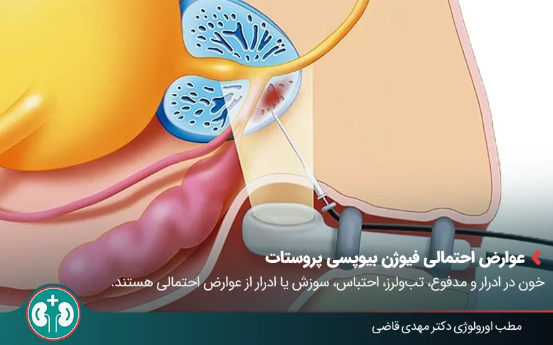
Possible Side Effects of Prostate Fusion Biopsy
Like any medical procedure, prostate fusion biopsy can have side effects and risks. These side effects are usually minor and manageable, and because this method is more accurate and targeted, they may be less common than with traditional biopsies. Awareness of these possible effects helps patients go through fusion biopsy with better preparation. The most common possible side effects include:
Bleeding: A small amount of blood in the urine, stool, or semen for a few days after fusion biopsy is normal. This usually resolves on its own. In rare cases, severe bleeding can occur.
Infection: After sampling, there is a risk of infection, especially in the rectum or urinary tract. For this reason, taking antibiotics before the procedure is important for prevention. Fever, chills, and severe pain can be signs of infection.
Urinary problems: Some patients may have burning with urination or frequent urination for a short time after biopsy. In rare cases, urinary retention, meaning inability to urinate, can also occur and requires evaluation by a urologist.
Cost and Access to Prostate Fusion Biopsy in Iran
The cost of prostate fusion biopsy in Iran can vary depending on the treatment center, insurance coverage, equipment used, and whether MRI fusion biopsy is performed at the same center.
In terms of access, this method is not yet common in all cities and treatment centers in Iran and is mostly available in specialized urology centers and clinics. Overall, although the initial cost of fusion biopsy is higher than traditional biopsy, its advantages in accuracy and reduced complications may make it a more cost-effective choice for many patients.
Summary
In this article, we discussed prostate fusion biopsy and compared it with traditional tissue sampling. We also explained its advantages and disadvantages compared with older methods and reviewed its side effects and cost. We hope this article has helped readers better understand this biopsy process.
In short, prostate fusion biopsy is a modern, advanced method for diagnosing prostate cancer. In this method, samples are taken in a targeted way from suspicious areas, which increases the chance of identifying clinically significant, higher-risk cancers and reduces the need for repeat biopsy.
Prostate fusion biopsy is a reliable and effective method for men who are at risk of prostate cancer or whose initial tests have been suspicious. If you are at risk of prostate cancer or would like to undergo the necessary examinations and evaluations, the best step is to see a urologist.
Frequently Asked Questions
Actions & related links
Related articles
All articlesWhat Is Polycystic Kidney Disease? (Fetal and Adult PKD)
What is polycystic kidney disease? Learn about inherited ADPKD and ARPKD, fetal and adult symptoms, complications, diagnosis, medicines, surgery, diet, fluids, and prevention-focused care.
What Is a Renal Cortical Cyst? Symptoms, Diagnosis, and Treatment
Renal cortical cyst | Simple vs. complex kidney cysts | Warning symptoms | Diagnosis and treatment | Needle drainage, laparoscopy, medication, and ablation
Kidney Transplant: Cost, Blood Type Compatibility, and Surgical Method
What is kidney transplant? Learn about cost considerations, operation duration and method, diet, who may not be eligible, success rates, isolation precautions, and post-transplant care.
What Is Pyelonephritis? Kidney Infection Symptoms, Diagnosis, and Treatment
What is pyelonephritis? Learn kidney infection symptoms in women, children, men, and pregnancy; diagnosis, antibiotics, treatment, emergency warning signs, and cystitis differences.
Comments
0 comments
No comments yet. Be the first to share your thoughts.