
 Tap to zoom
Urinary Diversion
Learn why urinary diversion is done, the main types, surgery options, stoma care, recovery, diet, activity, and follow-up needs.
- Published on
- June 26, 2026
- Reading time
- 5 min read
- Last updated
- Updated: June 27, 2026
The bladder and urinary control are not essential for survival, but they have a major effect on quality of life.
If, for any reason, there is an uncorrectable problem in the bladder’s storage or outlet system, or if other urinary pathways are blocked, there are ways to change the path of urine.
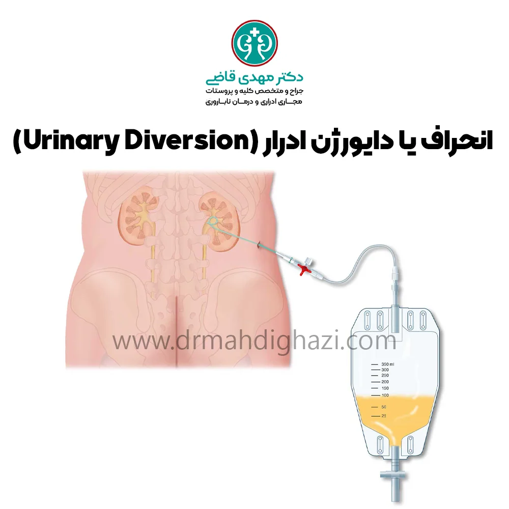
Urinary diversion surgery means redirecting the flow of urine for this purpose.
Normal function of the urinary system
The urinary tract consists of two kidneys, two ureters, the bladder, and the urethra. Urine is produced in the kidneys as waste products and water are removed, and it drains into the bladder through two tubes, 25 to 30 centimeters long, called ureters. The ureters are about six-tenths of a millimeter in diameter and have muscular walls that move urine toward the bladder.
The bladder can store urine until a person is ready to urinate. The ureteral valves or openings in the bladder also close the backward pathways toward the ureters so urine cannot return to the kidneys. The tube that carries urine out of the bladder is called the urethra.
Reasons for urinary diversion
Urinary diversion means changing the normal route of urine flow or creating a new route in the urinary system so urine exits through another pathway. Urinary diversion becomes necessary in the following situations:
Untreatable obstruction in the urinary system
The pathway through which urine is supposed to flow is no longer functioning as it did before and does not work adequately.
Urinary diversion may be needed in the following conditions:
Serious injury to bladder nerves, such as spinal cord injury, MS, spina bifida, and inability to urinate
Chronic bladder injury and irritation, such as interstitial cystitis
Causes that compress the ureter or urethra from outside
Trauma
Tumors
Large and complicated kidney stones
Types of urinary diversion
1. Bladder catheterization
Placement of a bladder catheter through the urethra
Placement of a bladder catheter through a small opening below the navel into the bladder, called suprapubic catheterization
2. Cystostomy
In the operating room, a catheter is surgically placed into the bladder through a small incision below the navel and fixed to the abdominal wall.
3. Nephrostomy
When the ureter is obstructed, a catheter is placed into the renal pelvis through a small opening by a surgeon or radiologist under imaging guidance, and the catheter remains in place for a defined period.
4. Ureteral stent
In cases of ureteral obstruction, if possible, using cystoscopy and under anesthesia, the surgeon passes the catheter through the urethra and bladder into the ureteral opening, opens the ureteral pathway, and advances it into the kidney. In some cases, even long-term stents, up to one year, may be used.
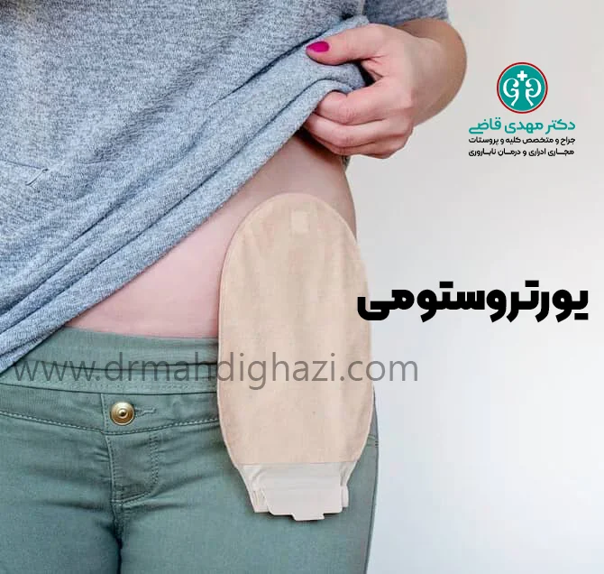
5. Ureterostomy
If the ureteral pathway is permanently closed, the ureter can be cut and its end connected to an opening the surgeon creates on the skin, so urine passes through the ureter and drains through the opening into a plastic urine bag attached to the ureterostomy opening. The patient empties this bag periodically.
6. Bladder diversion
This may be necessary if the bladder does not work properly or must be removed because of cancer or injury. In bladder urinary diversion, urine flow is redirected to a new bladder, called a neobladder, or routed outside the body through an opening in the abdominal wall, called a stoma.
How urinary diversion surgery is performed
Urinary diversion is divided into two broad types:
Continent urinary diversion
Incontinent urinary diversion
Incontinent Urinary Diversion
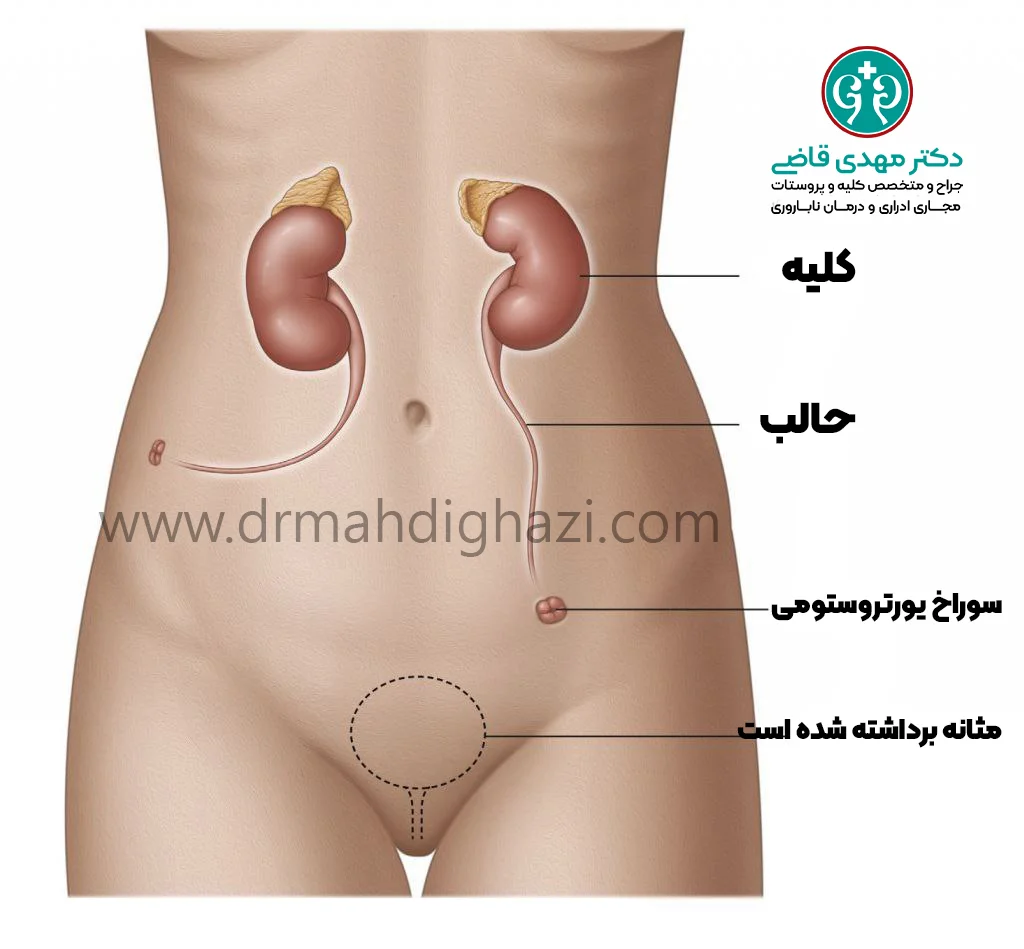
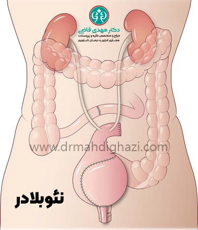
Ileal conduit: In this surgery, a new urine storage channel is created using intestine. One end opens onto the skin of the abdominal wall and the other end is connected to the ureters. Urine then flows from the ureters into this channel and continuously drains through its abdominal outlet into a plastic pouch called an ostomy bag, worn under clothing. When the plastic bag, or urostomy bag, becomes full, the person empties it.
Cutaneous ureterostomy: The surgeon diverts one of the ureters directly out of the body by connecting it to an opening on the skin of the flank, and a urine bag is fixed to the body over the skin opening.
Continent Urinary Diversion
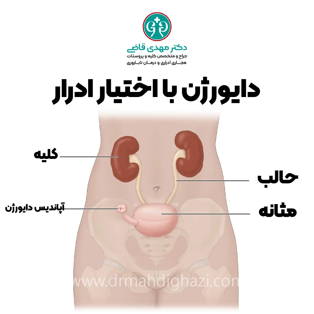
In continent urinary diversion, the surgeon creates a pouch from part of the intestine to hold urine inside the body. This urine reservoir has an outlet valve that prevents urine from leaking outside. One end of the pouch is connected to the ureters, and the other end is connected either to the urethra or to the abdominal skin. The bladder is removed from the body. There are two main types:
First type: It has an outlet valve made from the person’s appendix that exits through the abdomen, and the person must catheterize the urine reservoir several times a day through this valve to empty urine.
Second type: In this type, a neobladder, or new bladder, is created. With a neobladder, urination can be done in the usual way.
The advantage of both procedures is that there is no need to have a plastic urine bag on the abdomen. This can give people greater self-confidence and fewer activity limitations. One disadvantage of this method is the need for frequent daily catheterization.
After treatment
Most people who undergo urinary diversion are satisfied with the result and can return to normal life. However, problems can sometimes occur with the diversion, such as:
Changes in body fluid or salt levels
Difficulty inserting the catheter into the stoma
Problems related to skin overgrowth around the stoma and narrowing or blockage of the stoma
Serious surgical complications that may result from abdominal surgery, such as bowel obstruction or leakage of urine or stool
Postoperative care
After urinary diversion surgery, postoperative care plays a vital role in recovery and prevention of complications. The patient should spend the recovery period in the hospital so doctors can monitor their condition. During this time, pain control with pain medicines may be needed, and doctors can provide detailed instructions about medicines, nutrition, and permitted activities.
Care of the surgical site and stoma is very important. The patient should regularly keep the surgical site clean and dry to prevent infection. Regular dressing changes and stoma care may also be needed. Nurses and doctors provide the necessary training to the patient and family in this regard.
How to care for the stoma
The stoma, as a new pathway for urine to leave the body, requires special care. The skin around the stoma should always be kept clean and dry. Appropriate hygiene products should be used to prevent skin irritation and infection. In addition, regular replacement of the stoma bag and checking for any change in color or odor can help identify early problems.
The patient should learn how to empty and change the stoma bag correctly. This should be done carefully in a clean environment to prevent leakage or contamination. Special products such as skin-protective creams and powders can also help maintain the health of the skin around the stoma.
Proper nutrition and hydration after urinary diversion
Proper nutrition after urinary diversion surgery plays a key role in recovery. The patient should eat a diet rich in nutrients and fiber to support faster healing and prevent digestive problems. Adequate fluid intake is also very important to prevent dehydration and kidney stone formation.
Doctors may recommend that the patient avoid certain foods and drinks that can irritate the bladder or cause digestive problems. Eating low-fat, low-salt foods and avoiding processed foods can also support faster recovery.
Allowed physical activity and exercise after surgery
After urinary diversion surgery, patients should resume physical activity cautiously and in consultation with their doctor. In the first weeks after surgery, strenuous activity and lifting heavy objects should be avoided so the surgical site is not harmed.
Over time and with the doctor’s guidance, the patient can gradually increase physical activity.
Gentle exercise such as walking can improve blood circulation and speed recovery. However, strenuous sports and exercises that place significant pressure on the abdomen should be avoided until the doctor considers them appropriate.
Psychological and social support for patients
Psychological and social support is very important for patients who have undergone urinary diversion. Body changes after surgery can cause stress and anxiety. Therefore, access to psychological support, including counseling and support groups, can help the patient cope better with the new changes.
Family and friends also play an important role in supporting the patient. Providing emotional support and help with daily tasks can reduce the patient’s psychological burden. The patient can also benefit from the experiences and advice of others who have had similar experiences.
Ultimately, successful management of urinary diversion depends on a combination of appropriate medical care, proper nutrition and hydration, regular physical activity, and psychological and social support. Patients should stay in contact with their doctors and medical team and discuss any questions or concerns with them so they can find the best ways to manage their condition.
Summary
Urinary diversion is a surgical method used to change the normal pathway of urine flow in the body. It is usually necessary when the bladder or other urinary pathways can no longer function normally, such as in untreatable obstruction, chronic bladder diseases, trauma, tumors, and conditions such as spinal cord injury or neurologic diseases like MS and spina bifida.
Urinary diversion is divided into two broad types: continent and incontinent diversion. In incontinent methods such as ileal conduit and cutaneous ureterostomy, urine continuously drains into a plastic bag outside the body. In contrast, in continent diversion such as a neobladder, an internal reservoir is created to store urine, and the person must empty it regularly.
Postoperative care, especially care of the surgical site and stoma, plays a vital role in recovery and prevention of complications. Patients should keep the surgical site clean and dry and use appropriate hygiene products. Proper nutrition and hydration, avoiding heavy activity during recovery, and psychological counseling are also important after surgery.
Frequently asked questions
Actions & related links
Related articles
All articlesWhat Is Polycystic Kidney Disease? (Fetal and Adult PKD)
What is polycystic kidney disease? Learn about inherited ADPKD and ARPKD, fetal and adult symptoms, complications, diagnosis, medicines, surgery, diet, fluids, and prevention-focused care.
What Is a Renal Cortical Cyst? Symptoms, Diagnosis, and Treatment
Renal cortical cyst | Simple vs. complex kidney cysts | Warning symptoms | Diagnosis and treatment | Needle drainage, laparoscopy, medication, and ablation
Kidney Transplant: Cost, Blood Type Compatibility, and Surgical Method
What is kidney transplant? Learn about cost considerations, operation duration and method, diet, who may not be eligible, success rates, isolation precautions, and post-transplant care.
What Is Pyelonephritis? Kidney Infection Symptoms, Diagnosis, and Treatment
What is pyelonephritis? Learn kidney infection symptoms in women, children, men, and pregnancy; diagnosis, antibiotics, treatment, emergency warning signs, and cystitis differences.
Comments
2 comments