
 Tap to zoom
Ureterocele: Symptoms, Causes, Diagnosis, and Treatment
Ureterocele is a congenital abnormality often diagnosed in children under age two. Read about ureterocele symptoms, types, complications, diagnosis, and treatment options.
- Published on
- June 26, 2026
- Reading time
- 5 min read
- Last updated
- Updated: June 27, 2026
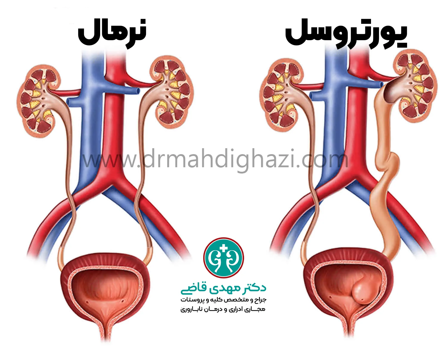
Most people are born with two ureters, one coming from each kidney and carrying urine to the bladder. In some people, however, about 1 in 500, two ureters come from one kidney. This is called ureteral duplication, or an extra ureter, and it can affect kidney health.
Ureteral Duplication and Ureterocele
Ureterocele is one of the conditions that can occur with ureteral duplication.
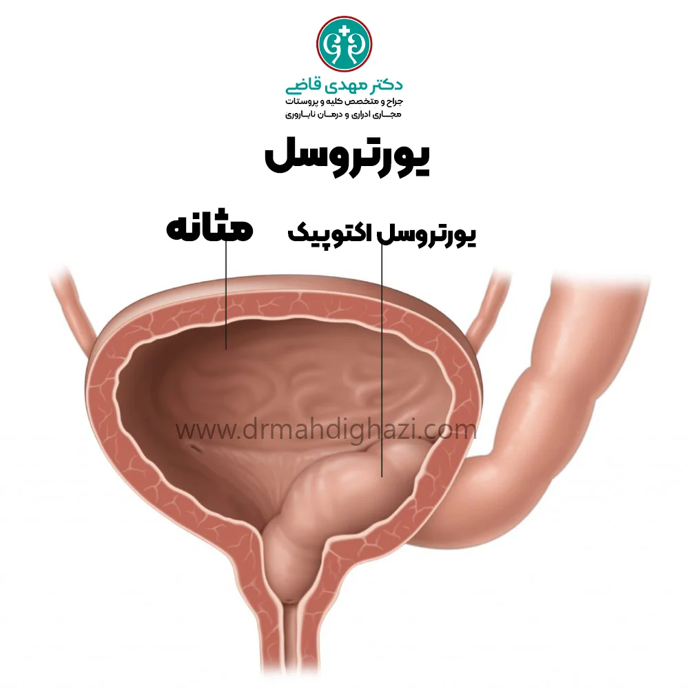
A ureterocele is a condition in which the ureter that connects the upper part of the kidney to the bladder becomes dilated and bulges into the bladder.
In ureteral duplication, the ureter that enters the bladder from the lower part of the kidney may also have reflux, meaning urine flows backward.
Ureterocele is a congenital abnormality that affects girls more often than boys.
Types of Ureterocele
A ureterocele means swelling at the end of the ureter, which can sit inside the bladder in a distinctive cobra-head shape.
In about 75% of cases, a ureterocele may form outside the bladder (the extravesical type), such as at the bladder neck or in the urethra, which can make diagnosis more difficult and time-consuming. In the intravesical type (25% of cases), this cobra-head-like swelling is seen on ultrasound or during bladder examination with a cystoscope.
If a person has two ureters in one kidney and the end of one of them forms a ureterocele, the second ureter may develop urinary reflux because the neighboring ureteral opening is often widened, allowing urine to flow back from the bladder to the kidney.
Fortunately, diagnostic tests and treatments can identify and correct this problem.
What Happens Normally?
The kidneys make urine by filtering waste products and excess water from the blood. Urine travels down from the kidneys through narrow tubes called ureters until it reaches the bladder, where urine is stored. After the bladder muscles contract, the stored urine leaves the body through the urethra.
There are one-way valves between the ureters and the bladder that move urine in only one direction. If urine mistakenly flows back toward the kidneys along this path, a condition called vesicoureteral reflux, or urinary reflux (VUR) may develop.
Different Forms of Ureterocele
Ureterocele can appear in different forms depending on the features below.
Degree of swelling
A ureterocele may become very swollen and take up a large amount of bladder space, or it may be only mildly swollen.
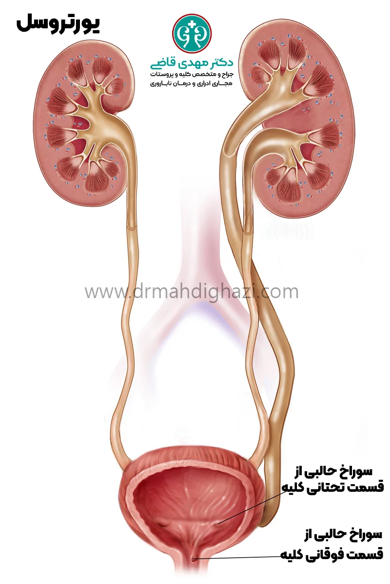
Location of the ureterocele
The end opening of the ureter can be in different locations: inside or outside the bladder, at the bladder neck, or in the urethra (outside the bladder, or ectopic).
Urinary reflux (VUR)
Ureterocele can occur with or without urine flowing back toward the kidneys (vesicoureteral reflux).
Ureterocele is most often found in children under two years old, but it may sometimes be seen in older children or adults. In some cases, it may even be diagnosed on pregnancy ultrasound while the fetus is still in the uterus.
Complications of Ureterocele
The main problems caused by ureterocele are kidney damage and kidney infection. Urinary obstruction can damage developing kidneys and reduce their ability to filter. Urine flowing back to the kidney (reflux) is also common, especially when there are two ureters in one kidney.
This happens because the ureterocele changes the normal shape of the one-way valves between the ureter and the bladder and affects how they work. In rare cases, reflux may even affect the opposite kidney.
There is also a small risk of kidney stone formation. Rarely, in girls, a ureterocele can protrude from the urethra and appear as a visible bulge.
Symptoms of Ureterocele
There are usually no symptoms. When symptoms are present, they may include:
Pain in the flank, back, or abdomen
Urinary tract infections, fever, and burning with urination
Foul-smelling urine
Frequent urination
Diagnosing Ureterocele
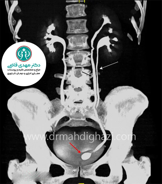
Ureterocele is often visible during pregnancy on maternal ultrasound. However, it may not be diagnosed until a child is evaluated for another problem, such as a urinary tract infection.
Ultrasound is the first imaging test used to look for this condition.
Other types of imaging may be done to understand the condition better or to help plan treatment.
For an infant or young child, the following tests may be performed:
Voiding cystourethrogram (VCUG)
This test is used to see how the bladder works. In this test, the bladder is filled with a special contrast dye, and X-ray images of the bladder and lower urinary tract are taken while the child urinates. First, a thin tube (catheter) is inserted through the urethra to fill the bladder with the contrast solution. Then, after the catheter is removed, several X-ray images are taken during urination. These images show the urologist problems with urine flow and abnormalities in the bladder's shape.
Nuclear kidney scan
After ureterocele is diagnosed, it is very important to check the kidneys for damage and for evidence of ureteral obstruction. A nuclear kidney scan shows these details.
Magnetic resonance imaging (MRI)
When the relevant anatomy is not clear enough, MRI may be done so the surgeon can be better prepared for possible surgery, if needed.
Treatment and Surgical Methods for Ureterocele
The timing and type of treatment are determined by several factors:
The patient's age and overall health
How the ureterocele affects kidney health
Whether urinary reflux is present
Sometimes more than one treatment method is needed. In uncomplicated cases, monitoring alone may be recommended. Treatment options include:
Surgery
Several types of surgery are used to treat ureterocele. The type of surgery depends on the location and severity of the ureterocele and on any associated problems.
Puncturing the ureterocele through the urethra (Transurethral Puncture)
In this method, the ureterocele is punctured with a cystoscope to lower its pressure. A cystoscope is a thin tube with a camera and a light at its tip.
This procedure usually takes 15 to 30 minutes and can be done without an overnight hospital stay.
This method does not require a large incision, but it may not be effective if the wall of the ureterocele is thick.
If it is not successful, open surgery may be needed.
This method works best when the ureterocele's outlet is inside the bladder.
Removal of the upper pole of the kidney or uretero-ureterostomy (Upper Pole Nephrectomy or Uretero-ureterostomy)
In some cases, the upper half of the kidney has stopped working because of the ureterocele. If there is no urinary reflux in the second ureter of the same kidney, the damaged part of the kidney may be removed.
This surgery is done through a small incision below the ribs or laparoscopically. Another option is used when the upper part of the kidney has not completely stopped working but is weak: its ureter can be connected to the second ureter of the same kidney.
This method is called uretero-ureterostomy and is performed through a small incision in the lower abdomen on the side of the ureterocele, or by laparoscopic or robotic surgery.
Nephrectomy, or kidney removal (Nephrectomy)
If the entire kidney does not work because of the ureterocele, it must be removed. This surgery can usually be performed laparoscopically or robotically.
Removal of the ureterocele and ureteral reimplantation into the bladder (Removal of the Ureterocele and Ureteral Reimplantation)
If removal of the ureterocele is necessary, surgery is performed.
For this surgery, the bladder is opened, the ureterocele is removed, the bladder floor and bladder neck are reconstructed, the ureter is reconnected to the bladder, and the ureteral valve is reconstructed to prevent urine from flowing back to the kidney.
This operation is done through a small incision in the lower abdomen.
This is a complex surgery, but it is successful in 90% to 95% of cases.
Ureteropyelostomy or upper-to-lower uretero-ureterostomy (Ureteropyelostomy or Upper-to-Lower Ureteroureterostomy)
If the upper part of the kidney works well and there is no reflux in the lower ureter, one option is to connect the blocked part to an unobstructed part of the ureter or kidney. This operation is done through a small incision in the lower abdomen. The success rate is 95%.
Antibiotics
Antibiotics are used to fight bacteria and help prevent kidney infection. In children who may have urinary obstruction or urinary reflux, a low-dose antibiotic may be prescribed daily until surgical correction to help prevent infection.
Frequently Asked Questions
Actions & related links
Related articles
All articlesWhat Is Polycystic Kidney Disease? (Fetal and Adult PKD)
What is polycystic kidney disease? Learn about inherited ADPKD and ARPKD, fetal and adult symptoms, complications, diagnosis, medicines, surgery, diet, fluids, and prevention-focused care.
What Is a Renal Cortical Cyst? Symptoms, Diagnosis, and Treatment
Renal cortical cyst | Simple vs. complex kidney cysts | Warning symptoms | Diagnosis and treatment | Needle drainage, laparoscopy, medication, and ablation
Kidney Transplant: Cost, Blood Type Compatibility, and Surgical Method
What is kidney transplant? Learn about cost considerations, operation duration and method, diet, who may not be eligible, success rates, isolation precautions, and post-transplant care.
What Is Pyelonephritis? Kidney Infection Symptoms, Diagnosis, and Treatment
What is pyelonephritis? Learn kidney infection symptoms in women, children, men, and pregnancy; diagnosis, antibiotics, treatment, emergency warning signs, and cystitis differences.
Comments
0 comments
No comments yet. Be the first to share your thoughts.