
 Tap to zoom
Ureteral Obstruction: Causes, Symptoms, Diagnosis, and Treatment
Learn how ureteral obstruction happens, warning symptoms, imaging tests, double-J stents, nephrostomy, surgery options, and kidney-protection timelines.
- Published on
- June 26, 2026
- Reading time
- 5 min read
- Last updated
- Updated: June 27, 2026
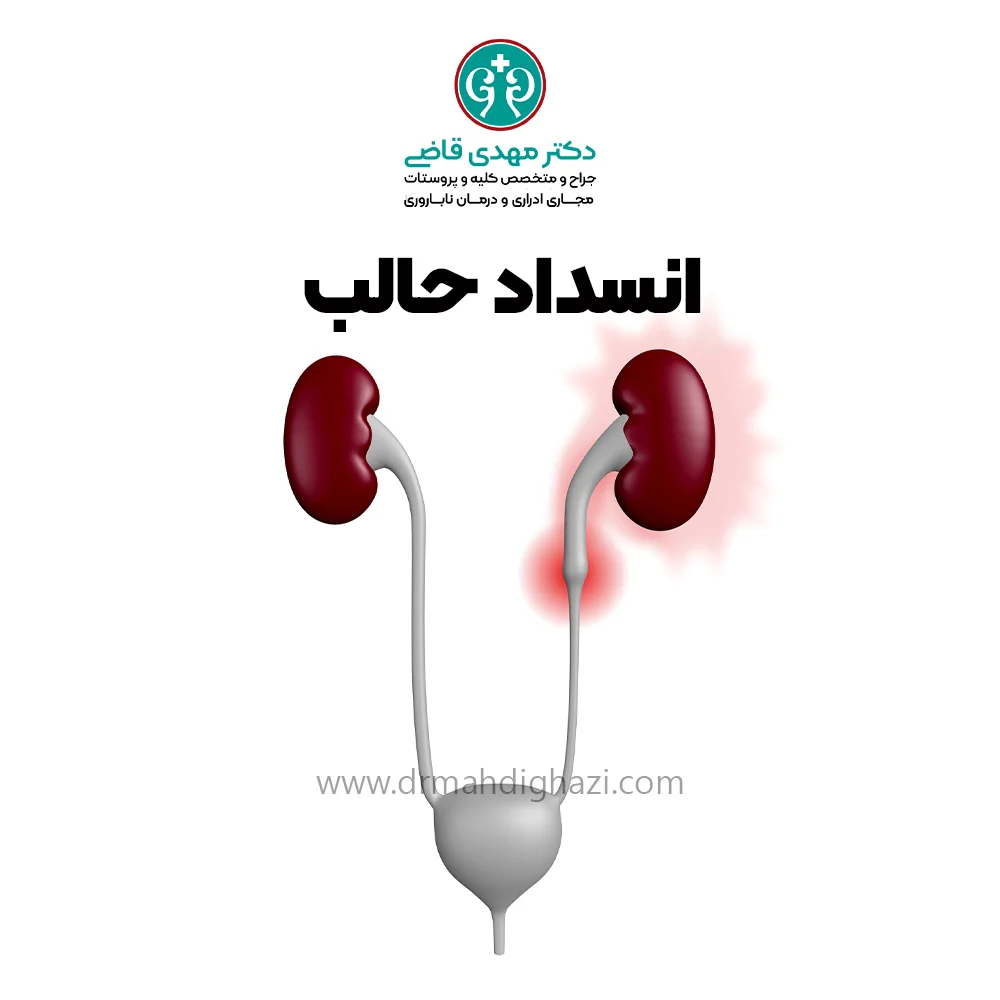
The ureter is a muscular tube that carries urine from the kidney to the bladder. It is about 25 centimeters long; its upper half is located in the abdomen and its lower half in the pelvis.
This urinary tube is about 3 millimeters in diameter and may become obstructed at different points along its course through the abdomen and pelvis.
If ureteral obstruction is not diagnosed and treated in time, it can lead to serious and even permanent consequences.
If complete ureteral obstruction remains persistent, after a period of time, usually two to three weeks, urine collects inside the kidney, the kidney becomes swollen, known as hydronephrosis, and permanent kidney damage may occur.
Ureteral obstruction can also cause high blood pressure, kidney failure, and an increased risk of urinary tract infections.
Internal obstruction occurs when the ureter becomes blocked because of causes such as narrowing, a ureteral stone, a blood clot, or masses.
External obstruction occurs when something outside the ureter presses on it and causes blockage.
Symptoms of external ureteral obstruction often develop gradually, and in mild cases there may be no symptoms.
However, if the obstruction is sudden and complete, the pain is usually severe.
In addition to pain, the following symptoms may occur:
Fever
Nausea
Vomiting
Difficulty passing urine
Bloody or dark urine
Causes of ureteral obstruction
Ureteral obstruction is an important problem in the urinary system and can occur for different reasons. It may be caused by internal factors such as stone formation, masses, and narrowing of the ureteral channel.
Internal factors can directly block the channel or disrupt normal ureter function by causing irritation and inflammation.
On the other hand, external pressure on the ureter from masses or structural changes around the channel can also cause obstruction.
These pressures may come from different areas of the abdomen and pelvis and interfere with the normal flow of urine.
Ureteral obstruction, whether caused by internal or external factors, can lead to urine buildup in the kidney and swelling of the kidney. If not treated in time, it may result in permanent kidney damage.
Internal causes of obstruction include:
Ureteral stone
Ureteral masses, polyps, and tumors
Blood clot
Tuberculosis of the ureter and schistosomiasis
Ureterocele
Congenital ureteral stricture
Injury caused by endoscopic surgical instruments
External causes of ureteral obstruction block the ureter by pressing on it and include:
Benign and malignant tumors of the abdomen and pelvis
Vascular disorders and masses of large blood vessels, such as aortic aneurysm
Noncancerous conditions in women, including pregnancy, uterine or ovarian masses, Gartner duct cyst, endometriosis, uterine prolapse, and ureteral injury during uterine or ovarian surgery
Gastrointestinal diseases, including Crohn’s disease, appendiceal inflammation, diverticulitis, and pancreatic lesions
Other large abdominal masses
Retroperitoneal fibrosis
Several factors can increase the risk of ureteral obstruction.
These include a history of abdominal or pelvic surgery, radiation therapy to the pelvis, chronic urinary tract infections, and congenital abnormalities of the urinary system.
Use of certain medicines, such as anticholinergics, which can reduce urine movement, may also increase the risk of ureteral obstruction.
Diagnostic methods
Temporary ureteral obstruction is very common, and the most common cause is kidney stones that are passing. Imaging is the key to diagnosing ureteral obstruction, and any of the following imaging tests may be performed:
Ultrasound
CT scan
MRI
IVP, an X-ray study with contrast material
Nuclear kidney scan
CT scan is an excellent imaging method for detecting ureteral stones, while IVP provides a more detailed image of the ureter’s structure, shape, and anatomy.
These tests often show the location of the obstruction and may also identify its exact cause.
Urine and blood tests are also used to assess kidney function.
Treatment options
Treatment usually depends on the type of obstruction. The first emergency step is to establish adequate drainage of urine from the kidneys so the pressure caused by obstruction is relieved and the risk of kidney damage is reduced.
This can be done with the following methods:
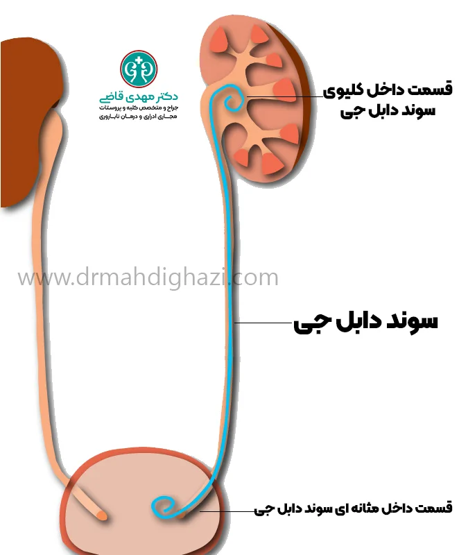
Ureteral stent or double-J catheter
A thin, hollow plastic tube is usually placed under anesthesia through the urinary tract between the kidney and bladder inside the ureter, temporarily keeping the ureter open for urine flow for several months.
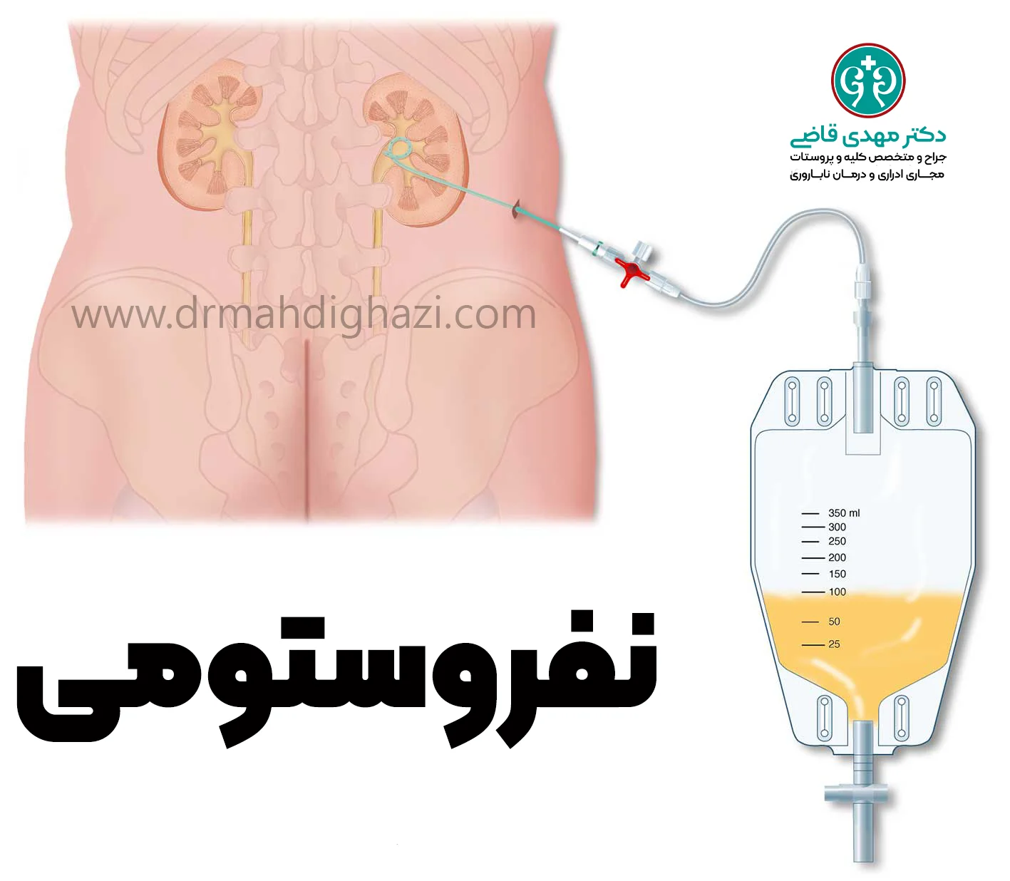
Nephrostomy
In this method, a catheter, guided by X-ray or ultrasound under local anesthesia or general anesthesia, is passed through the skin of the back into the kidney so urine inside the kidney can drain out of the body. The end of this catheter is connected to a drainable plastic bag.
After the obstruction has been relieved, laparoscopic surgery or open surgery may be needed to correct the obstruction permanently.
The type of surgery differs depending on the cause of obstruction. If a mass from outside is pressing on the ureter, surgery is performed to remove the mass.
If narrowing is the cause of obstruction, the narrowed part of the ureter may be surgically removed.
Some obstructive causes inside the ureter, such as stones, are treated with ureteroscopy, which is endoscopy of the ureter.
In cases where the cause of obstruction cannot be removed, the urologist must divert the ureteral pathway to another location where there is no obstruction.
Usually, one of the following methods is used for urinary diversion (urinary diversion):
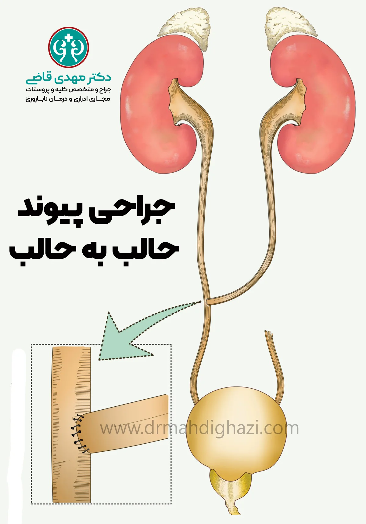
In the first method, the ureters are connected to each other so the normal urine pathway from one kidney to the other ureter is established and urine drains through it into the bladder. This is ureter-to-ureter anastomosis, or ureteroureterostomy.
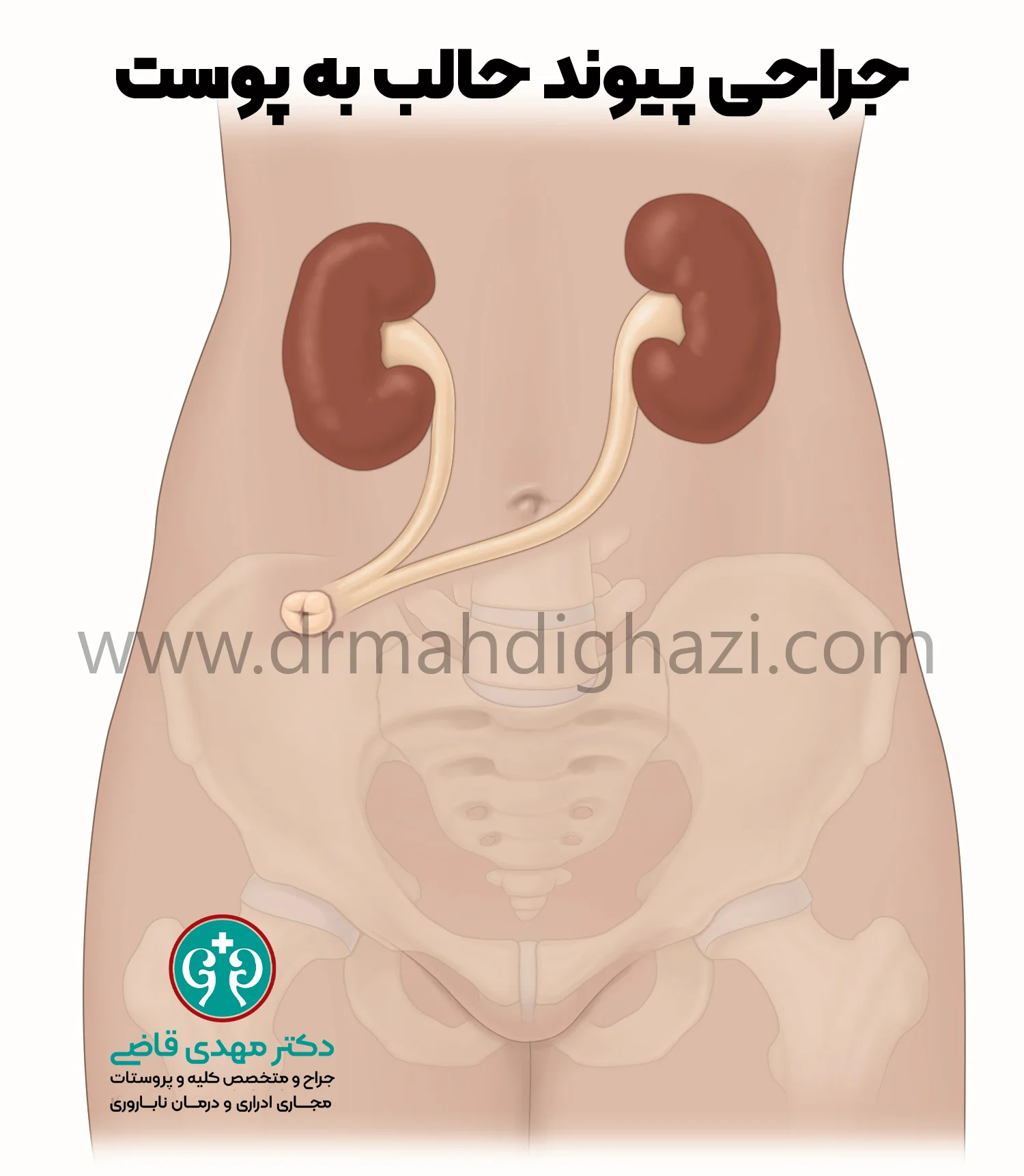
In the second method, the ureter is cut and its end is connected to an opening on the abdomen. Through this opening, urine passes outside the body into a drainable plastic bag. This is ureter-to-skin anastomosis, or ureterostomy.
After treatment
Outcomes can vary.
Any kidney injury caused by long-term obstruction can lead to permanent damage.
However, if the cause of obstruction is diagnosed early and corrected, kidney damage is usually minimal and negligible.
If the kidney is completely obstructed for more than two months and its urinary pathway is not restored, it will usually stop functioning completely.
In general, if only one kidney is affected, fortunately the other kidney often enlarges in compensation and tries to take over some of the work of the unhealthy kidney.
Frequently asked questions
Actions & related links
Related articles
All articlesWhat Is Polycystic Kidney Disease? (Fetal and Adult PKD)
What is polycystic kidney disease? Learn about inherited ADPKD and ARPKD, fetal and adult symptoms, complications, diagnosis, medicines, surgery, diet, fluids, and prevention-focused care.
What Is a Renal Cortical Cyst? Symptoms, Diagnosis, and Treatment
Renal cortical cyst | Simple vs. complex kidney cysts | Warning symptoms | Diagnosis and treatment | Needle drainage, laparoscopy, medication, and ablation
Kidney Transplant: Cost, Blood Type Compatibility, and Surgical Method
What is kidney transplant? Learn about cost considerations, operation duration and method, diet, who may not be eligible, success rates, isolation precautions, and post-transplant care.
What Is Pyelonephritis? Kidney Infection Symptoms, Diagnosis, and Treatment
What is pyelonephritis? Learn kidney infection symptoms in women, children, men, and pregnancy; diagnosis, antibiotics, treatment, emergency warning signs, and cystitis differences.
Comments
2 comments