
 Tap to zoom
What Is Transrectal Prostate Ultrasound and Why Is It Done?
What is transrectal prostate ultrasound? | Uses | Benefits and limitations | Preparation before TRUS | Procedure steps and duration | Aftercare | Is it risky?
- Published on
- June 26, 2026
- Reading time
- 5 min read
- Last updated
- Updated: June 27, 2026
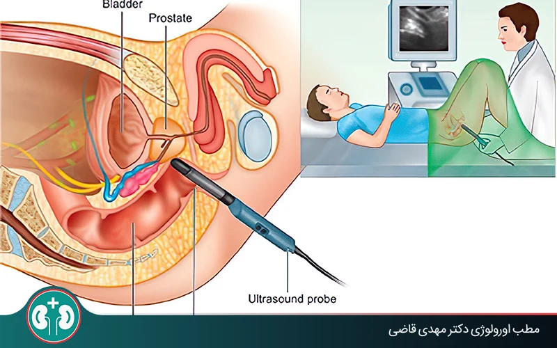
Transrectal prostate ultrasound is a precise, minimally invasive form of prostate ultrasound that urologists use to evaluate different problems of the prostate gland. In this procedure, a small probe is inserted into the rectum and uses sound waves to create clear, real-time images of the prostate.
In this article, we explain in detail what transrectal prostate ultrasound is, when it is performed, and how it can help diagnose conditions such as inflammation, benign enlargement, or even prostate cancer. We also discuss its benefits, limitations, care before and after the test, and newer technologies used with this method.
Urologic surgeon Prostate disease specialist
Note: To improve care quality and patient satisfaction, appointments are triaged by reason for visit. Each physician sees patients within the relevant urology subspecialty.
Book appointment
If you are looking for a practical, comprehensive guide to transrectal ultrasound, keep reading.
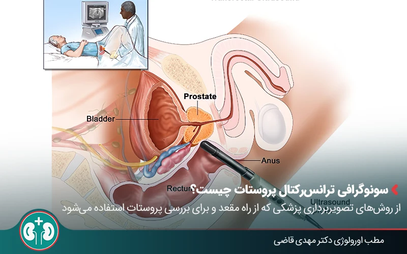
What Is Transrectal Prostate Ultrasound?
Transrectal ultrasound (TRUS) is a common medical imaging method used especially to examine the prostate. Urologists use prostate ultrasound to diagnose problems such as prostate enlargement or suspicious masses.
The main purpose of transrectal prostate ultrasound is to obtain a clear image of prostate gland tissue so any problem can be detected earlier. A small device is inserted through the rectum and uses sound waves to create a detailed image of the prostate.
Appointments related to Urologic surgeon Prostate disease specialist
Open the public booking path to review times and related information.
Book appointment
New Methods in Transrectal Prostate Ultrasound
In recent years, transrectal prostate ultrasound has advanced considerably. These advances help doctors make decisions about prostate treatment or surgeries such as prostatectomy, which means complete removal of the prostate, with greater confidence.
Below are newer methods and technologies used in transrectal prostate ultrasound:
Contrast-Enhanced Ultrasound
In this method, a substance called a contrast agent is injected so blood vessels can be seen more clearly on ultrasound images. Some cancerous tissues form more blood vessels, and in these cases contrast-enhanced ultrasound can show areas suspicious for cancer more clearly.
Q&A — Urologic surgeon Prostate disease specialist
General questions are shown on the destination page after review.
Book appointment
Elastography
Elastography is a newer technology that measures how firm or soft tissues are. Because tumors are usually firmer than healthy tissue, this difference can be seen with a special ultrasound technique called elastography. In other words, this method can show suspicious areas more clearly than ordinary ultrasound.
Combined MRI and Ultrasound Imaging
This method, also called "prostate fusion biopsy", uses images from MRI and transrectal ultrasound at the same time. It has very high diagnostic accuracy and is especially useful for patients whose previous biopsy was negative but whose prostate-specific antigen (PSA) level remains high.
To perform it, MRI is first used to identify suspicious areas in the prostate. Those same areas are then located with live ultrasound imaging, and targeted biopsy samples are taken from them.
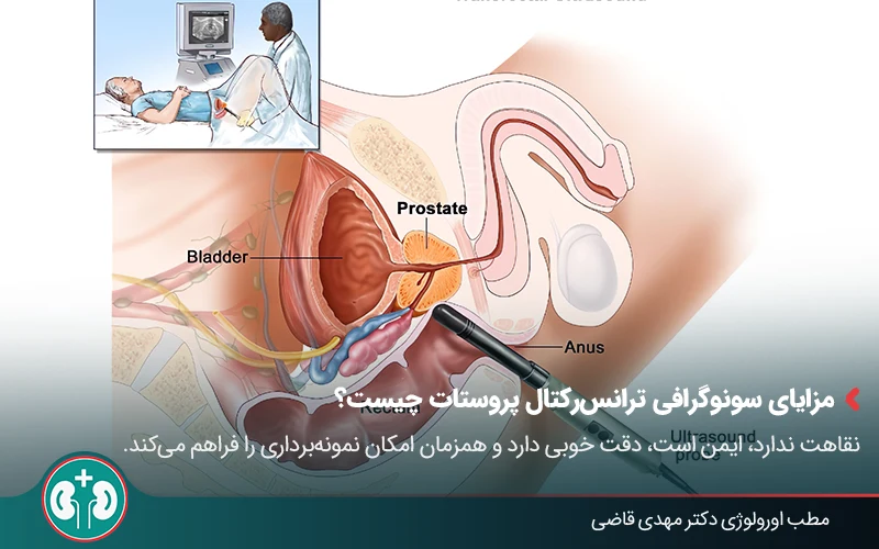
Benefits and Limitations of Transrectal Ultrasound
Compared with other imaging methods, transrectal ultrasound (TRUS) is a safe procedure; however, it also has limitations, so the doctor may sometimes need other diagnostic methods for a more detailed evaluation. Below, we review its benefits and limitations.
Benefits
This method is useful in diagnosing and evaluating the prostate because of the following features:
High safety: Unlike CT scans or X-rays, transrectal ultrasound (TRUS) uses sound waves only and does not use ionizing radiation. Therefore, it is considered safe, and repeating it is not harmful.
Coverage accuracy:Because the prostate sits deep in the pelvis, this method allows accurate visualization of areas that cannot be felt on digital rectal examination, such as the base and apex of the prostate.
Ability to perform procedures at the same time: If needed, tissue sampling (biopsy) can be performed at the same time.
Ease of performance: It does not require hospitalization and is completed in a short time, usually 15 to 30 minutes.
Limitations
Despite these advantages, this method also has challenges:
Patient discomfort: Insertion of the probe through the rectum may be uncomfortable for some people.
Diagnostic limitation: It has limitations in detecting very small cancers or early-stage disease, and additional tools such as MRI or PSA testing are often needed.
Uses of Transrectal Prostate Ultrasound
Transrectal ultrasound is used not only in prostate cancer diagnosis, but also in evaluating benign enlargement, structural abnormalities, and even fertility problems. Below, we review the uses of transrectal prostate ultrasound in more detail.
1. Diagnosis of Prostate Cancer
Transrectal ultrasound helps the urologist identify areas inside the prostate that are suspicious for cancer. Tumors often appear as dark areas on these images. If an area is suspicious, a biopsy (tissue sample) is taken from that same area.
2. Identifying Benign Prostatic Hyperplasia (BPH)
With this ultrasound, the prostate size can be measured accurately, and changes related to natural prostate enlargement (BPH) can be identified.
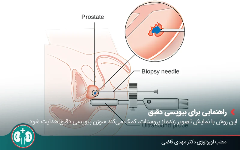
3. Guidance for Accurate Biopsy or Local Treatments for Prostate Cancer
If the specialist needs to take a prostate biopsy, knowing the tumor location allows more accurate sampling. Treatments such as freezing the tumor (cryotherapy) or applying high-intensity focused ultrasound (HIFU) for prostate cancer also require precise guidance.
Transrectal ultrasound acts like the specialist's eyes and shows the exact location of the tumor. By providing live, real-time images from inside the prostate, it also helps guide the biopsy needle or treatment instruments exactly to the intended location.
4. Evaluating the Cause of Male Infertility (Azoospermia)
If a man has fertility problems and the doctor suspects obstruction of the ejaculatory ducts, this ultrasound can evaluate the sperm outflow pathway and the seminal vesicles.
When Is Transrectal Prostate Ultrasound Needed?
Specialists usually recommend transrectal prostate ultrasound when there is evidence of abnormal changes or suspicious symptoms in the body.
The following are situations in which this ultrasound may be needed for prostate disease:
A firm or abnormal area found during digital rectal examination of the prostate;
A high prostate-specific antigen (PSA) level on a blood test;
Presence of blood in semen or urine;
Suspicion of benign prostatic hyperplasia based on symptoms or test findings;
Need to monitor treatment progress in patients with prostate diseases.
Preparation Before Transrectal Prostate Ultrasound
To help transrectal prostate ultrasound be performed correctly and without problems, it is best to follow a few simple steps before your appointment:
Wash the genital and anal area with soap and water so the body is completely clean.
Prepare a list of the medicines you take and provide it to the urologist if needed. If you take blood thinners such as aspirin or warfarin, be sure to ask your doctor whether you should stop or continue them. This point is very important for biopsy.
Be sure to consult your urologist before the test to make sure you have the necessary preparation.
Steps of Transrectal Prostate Ultrasound
Although transrectal prostate ultrasound may feel a little worrying, the process is simple, brief, and tolerable. Below are the steps so you know what to expect and what will be done:
Preparation
On the day of your appointment, after entering the imaging department, you may be asked to wear a hospital gown. In some cases, you may need to empty your bladder before the procedure begins.
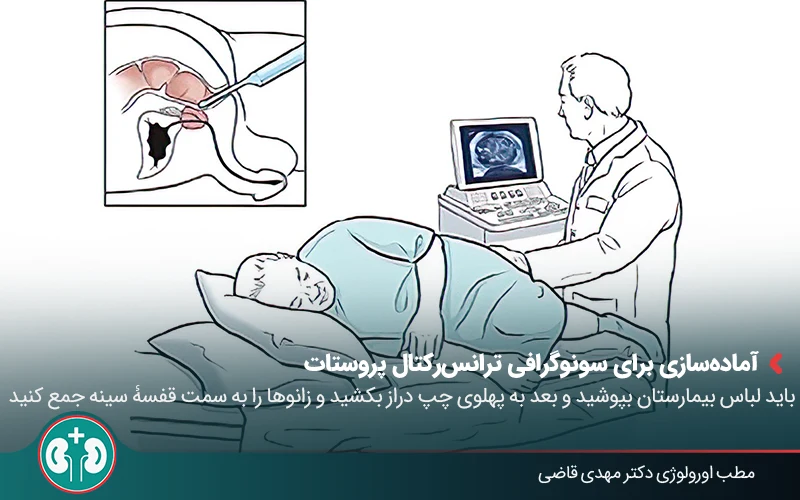
Before the ultrasound starts, the radiologist asks you to lie on your left side and bring your knees toward your chest. This position places the prostate in the best position for imaging. The doctor or technician then covers the ultrasound probe with a protective sheath and applies a special gel. The gel helps the probe enter the rectum more easily.
Performing the Transrectal Ultrasound and Biopsy if Needed
The radiologist gently places the probe inside the rectum. At this moment, you may feel some pressure or discomfort, but this feeling is usually brief and tolerable. Images appear live on the monitor, and the radiologist examines the shape, size, and internal structure of the prostate. If a suspicious area is seen, a biopsy is also performed at the same time.
For the biopsy, the specialist first injects a local anesthetic. Then a thin needle is placed alongside the probe, and several small samples of prostate tissue are taken.
Immediately After Transrectal Ultrasound
After the examination or biopsy is finished, the radiologist removes the probe and you can get dressed. If you had ultrasound only, you can return to your daily activities immediately. If a biopsy was also performed, the urologist will explain the necessary instructions.
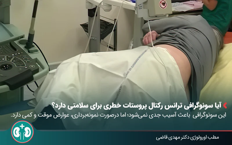
Is Transrectal Prostate Ultrasound Risky for Health?
This ultrasound by itself does not cause any serious injury or risk, and there is usually no need to worry. However, if tissue sampling from the prostate, or biopsy, is performed at the same time, some temporary and manageable symptoms may occur:
Blood in semen and urine;
Pain or discomfort in the pelvis or rectum;
Pain or burning during urination;
Difficulty passing urine;
Mild bleeding from the rectum;
Possibility of a mild infection.
These effects are actually side effects of prostate biopsy and are usually not severe and resolve on their own within a few days.
Aftercare Following Transrectal Prostate Ultrasound
Transrectal prostate ultrasound alone does not require special care, but if it is performed with biopsy, self-care and following these simple points are important:
1. Drink fluids: In the first days after biopsy, drink plenty of fluids, especially water. This can help reduce burning during urination and help clear possible blood or clots more quickly.
2. Manage medicines:Take prescribed antibiotics and pain relievers exactly as your doctor instructs. Restart medicines such as aspirin or ibuprofen only if your urologist approves.
3. Limit physical activity: Avoid strenuous exercise, heavy lifting, and high-pressure physical activity for 24 to 48 hours after biopsy, especially if you still see bright red blood in the urine.
4. Delay sexual intercourse: Do not have sexual intercourse until you feel comfortable.
5. Contact your doctor:Pay attention to any sudden change in urination, inability to pass urine, high fever, severe pain, or unusual bleeding. If these symptoms occur, contact your doctor immediately.
Summary
Transrectal prostate ultrasound is a minimally invasive and safe method for evaluating the prostate gland, and it plays an important role in diagnosing problems such as benign enlargement, tumors, and even male infertility.
Because TRUS provides a complete image of the prostate, does not use radiation, is quick to perform, and can be combined with advanced methods such as MRI, it is an important tool for diagnosing and monitoring prostate diseases.
This prostate imaging method is performed by inserting a thin probe into the rectum and, when necessary, allows accurate biopsy of suspicious areas.
Men over age 50, or those with symptoms such as frequent urination, pelvic pain, or a high PSA level, may be candidates for this imaging and should see a urologist. The doctor can decide whether transrectal ultrasound is needed based on symptoms, family history, and test results.
Frequently Asked Questions
Sources:
Actions & related links
Related articles
All articlesWhat Is Polycystic Kidney Disease? (Fetal and Adult PKD)
What is polycystic kidney disease? Learn about inherited ADPKD and ARPKD, fetal and adult symptoms, complications, diagnosis, medicines, surgery, diet, fluids, and prevention-focused care.
What Is a Renal Cortical Cyst? Symptoms, Diagnosis, and Treatment
Renal cortical cyst | Simple vs. complex kidney cysts | Warning symptoms | Diagnosis and treatment | Needle drainage, laparoscopy, medication, and ablation
Kidney Transplant: Cost, Blood Type Compatibility, and Surgical Method
What is kidney transplant? Learn about cost considerations, operation duration and method, diet, who may not be eligible, success rates, isolation precautions, and post-transplant care.
What Is Pyelonephritis? Kidney Infection Symptoms, Diagnosis, and Treatment
What is pyelonephritis? Learn kidney infection symptoms in women, children, men, and pregnancy; diagnosis, antibiotics, treatment, emergency warning signs, and cystitis differences.