
 Tap to zoom
Megaureter: Urinary Obstruction with a Dilated Ureter
Learn what megaureter is, its types, urinary infection symptoms, ultrasound, VCUG, renal scan, MRI, surgery, and follow-up for kidney protection.
- Published on
- June 26, 2026
- Reading time
- 5 min read
- Last updated
- Updated: June 26, 2026
Most children are born with a normal urinary tract, but some infants develop a condition called megaureter. In this condition, the tube that connects the kidney to the bladder becomes wider and more dilated than usual. This problem can cause infection and obstruction of urine flow, and if it is not treated, it may cause serious kidney damage.
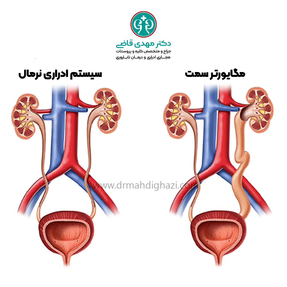
Definition of Megaureter
Megaureter is a condition in which the ureter, the tube that connects the kidney to the bladder, becomes wider than 1 centimeter. This ureteral dilation can happen for two reasons: 1. a congenital abnormality in the structure of the ureter, called primary megaureter; 2. backflow of urine from the bladder into the ureters, called secondary megaureter.
Types of Megaureter
Megaureter is divided into four main types, each with its own features and problems:
Primary Obstructive Megaureter
In this type, there is an obstruction where the ureter enters the bladder. This increases pressure in the ureter and causes it to dilate, and over time this obstruction can damage the kidney. Surgery may be needed to correct this problem.
Follow-up for this type of megaureter is very important even when there are no symptoms.
Refluxing Megaureter
This type of megaureter is caused by urine flowing backward from the bladder into the ureters. Normally, urine should not flow back from the bladder into the ureters, but in this type of megaureter, high bladder pressure causes urine to reflux into the ureters. This type is seen more often in male infants. Sometimes the problem improves on its own during the first year of life, but if it does not improve, surgery may be needed.
Nonobstructive, Nonrefluxing Megaureter
In this type, the ureters are wider than usual without any obvious reason. In other words, there is no obstruction and no urinary reflux. This type of megaureter is congenital and improves over time in many cases. The urologist must make sure there is no obstruction or urinary reflux in these patients.
Obstructive and Refluxing Megaureter
This type of megaureter combines obstruction and urinary reflux. It is dangerous because the ureters gradually become more dilated and a more severe obstruction develops. These patients are at greater risk of urinary tract infections.
Secondary Megaureter
This type of megaureter develops because of other diseases. Some abnormalities that can cause megaureter include:
Posterior urethral valves, an obstruction in the male urethra
Prune-Belly syndrome
Neurogenic bladder, meaning lack of normal bladder control because of neurologic problems such as spina bifida or spinal cord injury
Symptoms of Megaureter
Doctors usually diagnose megaureter while evaluating children who have a urinary tract infection.
Today, because prenatal ultrasound is widely used, many megaureters are identified before birth when hydronephrosis, or kidney dilation, is seen in the fetus.
Methods for Diagnosing Megaureter
To diagnose megaureter, urologists use several tests and imaging methods to evaluate urinary tract function. These tests include:
Ultrasound
This painless, noninvasive test is used to evaluate the appearance of the kidneys, ureters, and bladder. Ultrasound is very effective for finding dilated ureters and provides detailed images of dilated ureters in megaureter.
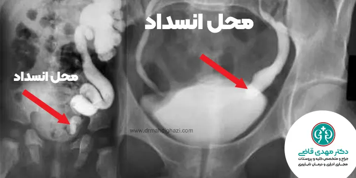
Voiding cystourethrography, or VCUG
This X-ray test is used to check for urine reflux into the ureters.
In this method, a small catheter is inserted through the urethra into the bladder, and a special contrast material is injected into the bladder. Then, during urination, X-ray images are taken to show whether urinary reflux is present.
Nuclear kidney scan with diuretic medicines
This scan is used to evaluate ureteral obstruction.
In this test, a small amount of radioactive fluid is injected into a vein. It travels through the bloodstream to the kidneys and is then excreted in the urine.
This test shows how each kidney is working and whether there is an obstruction in the ureter.
MRI of the urinary tract, MR-U
This imaging method uses magnetic fields to create detailed images inside the body.
MRI shows the structure of the urinary tract better than ultrasound or a nuclear scan.
This method is usually not performed in young children because it requires sedation or general anesthesia.
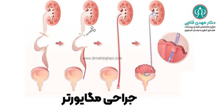
Open Surgical Treatment of Megaureter
If evaluation shows obstruction or reduced kidney function, the child may need surgery.
In standard surgery, the ureter is separated from the bladder, its end is narrowed, and it is reattached to the bladder in another location.
If the child does not have a urinary tract infection or reduced kidney function, surgery can be delayed until 1-2 years of age.
Surgery in infants should be performed by surgeons experienced in pediatric surgery, and many infants receive antibiotic treatment until surgery to prevent infections.
Final Points
Megaureter is a serious problem that requires timely diagnosis and treatment.
With appropriate diagnostic tests and treatments, many children with megaureter can improve and avoid serious complications.
The most important point is that parents should see a doctor promptly if they notice any suspicious symptoms and should complete the necessary follow-up.
Frequently Asked Questions
Actions & related links
Related articles
All articlesWhat Is Polycystic Kidney Disease? (Fetal and Adult PKD)
What is polycystic kidney disease? Learn about inherited ADPKD and ARPKD, fetal and adult symptoms, complications, diagnosis, medicines, surgery, diet, fluids, and prevention-focused care.
What Is a Renal Cortical Cyst? Symptoms, Diagnosis, and Treatment
Renal cortical cyst | Simple vs. complex kidney cysts | Warning symptoms | Diagnosis and treatment | Needle drainage, laparoscopy, medication, and ablation
Kidney Transplant: Cost, Blood Type Compatibility, and Surgical Method
What is kidney transplant? Learn about cost considerations, operation duration and method, diet, who may not be eligible, success rates, isolation precautions, and post-transplant care.
What Is Pyelonephritis? Kidney Infection Symptoms, Diagnosis, and Treatment
What is pyelonephritis? Learn kidney infection symptoms in women, children, men, and pregnancy; diagnosis, antibiotics, treatment, emergency warning signs, and cystitis differences.
Comments
0 comments
No comments yet. Be the first to share your thoughts.