
 Tap to zoom
What Is Cystoscopy?
Cystoscopy lets a urologist examine the bladder through the urethra. Learn why it is done, how flexible and rigid cystoscopy work, recovery tips, and warning symptoms.
- Published on
- June 26, 2026
- Reading time
- 5 min read
- Last updated
- Updated: June 26, 2026
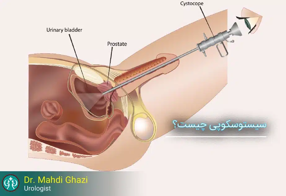
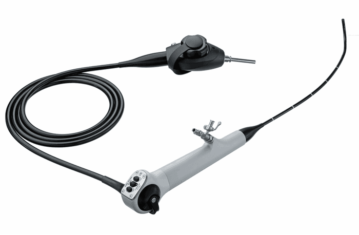
Cystoscopy, or cysto-urethroscopy, is a procedure in which a urologic surgeon (urologist) can use a cystoscope to examine the patient's bladder in precise detail through the urethra (the urine outlet channel). Cystoscopes have a lens and a light source; some have additional capabilities and can assist during surgical procedures.
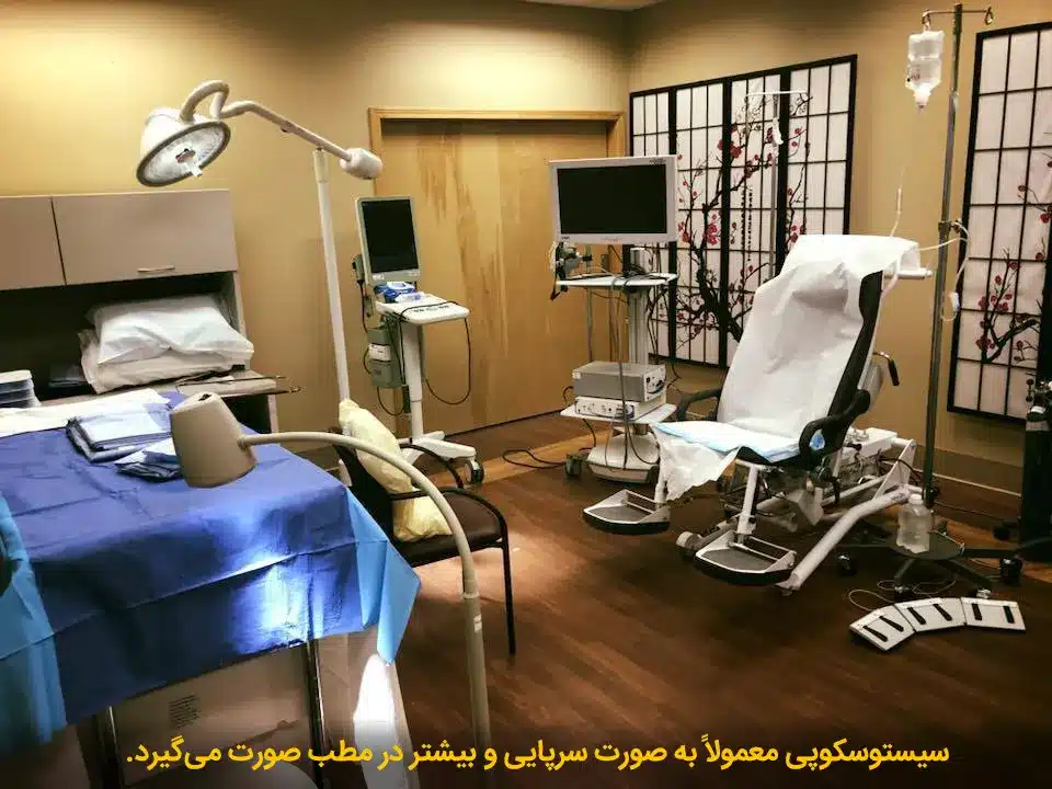
Cystoscopy is performed with local anesthesia and takes about 5 to 10 minutes (in the urologist's office).
Reasons for Cystoscopy
This method is often used to diagnose the cause of the following problems:
Obstruction in the urinary tract
Bladder abnormalities or abnormalities of its lining, such as a tumor or growth
How Is Cystoscopy Performed?
Before the procedure:
Before cystoscopy, the patient should remove all outerwear and underwear and put on an operating-room gown. The patient then lies on the bed in a position with the legs apart so the physician can access the urinary tract. To prevent pain, a special gel is used to provide local anesthesia of the urethra in the urinary tract. General or spinal anesthesia is usually not used. After anesthesia is achieved, the physician inserts the thin cystoscope tube into the urethra and examines the inside of the bladder and the passages.
Because urinary tract infection is a common problem after cystoscopy surgery, the physician may, for prevention, ask the patient to have a urinary tract infection test before the procedure or to take a course of antibiotics before the procedure. During the operation, general anesthesia is also usually used. It is essential for a close companion to accompany the patient after surgery, and the patient should not personally drive after the operation.
During the procedure:
The patient will lie on the examination table. An anesthetic gel is injected into the urethra using a syringe without a needle tip. The patient may receive a small amount of sedative medication through the veins (intravenously).
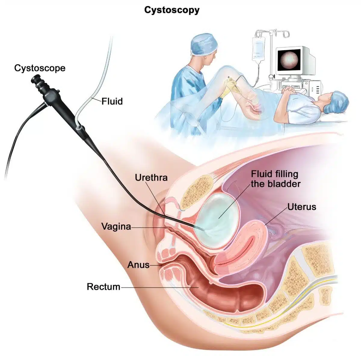
The cystoscope (endoscope), a flexible catheter-like camera equipped with a light and lens, is inserted into the bladder through the urethra. The entire length of the urethra and the inside of the bladder can be evaluated by cystoscopy. During cystoscopy, a sterile saline solution is introduced into the bladder to give the urologic surgeon a better view. Cystoscopy takes about 5 to 10 minutes on average.
What Should You Do After Cystoscopy?
Although most people recover within one or two days after the procedure, for faster recovery it is best to keep the following points in mind:
Rest if you feel tired. Getting enough sleep is one of the things that helps your recovery.
Avoid strenuous or heavy activity until your physician approves it.
Ask your physician when you can start driving and resume sexual activity.
You can continue your regular daily diet. Drinking fluids is also helpful.
Take your medications exactly as prescribed by your physician. If you have stomach pain, take the medicine after eating.
Be sure to see your physician on the scheduled date and complete the full course of antibiotics.
Have the necessary tests done and show the results to your specialist.
A small amount of blood in the semen after cystoscopy is normal; however, if the patient has severe pain, fever, or significant bleeding after cystoscopy, the physician should be informed as soon as possible.
Another type of cystoscope is metallic and is used under anesthesia in the operating room. Larger instruments can be passed into the bladder through this type of cystoscope, giving the surgeon more ability to perform small operations on the bladder.
Additional notes:
During cystoscopy, the surgeon may use small instruments that enter the bladder through the endoscope and can be used to take a biopsy or remove small growths.
For one or two days after the procedure, the patient may have mild burning or discomfort when urinating.
After cystoscopy, the bladder wall should appear smooth, and the size, shape, and position of the bladder should be normal. There should also be no obstruction or blockage.
Types of Cystoscopy and How They Are Performed
The type of cystoscope the specialist uses depends on the purpose of the examination. The cystoscopy procedure is performed using an instrument called a cystoscope. At the end of this instrument, which is a thin tube, there is a small light. Two types of cystoscopy procedure can be named:
Rigid or standard rigid cystoscope
Flexible cystoscope
Rigid Cystoscope
Because the cystoscope tube has a large diameter, rigid cystoscopy is not performed using local anesthesia; because it is painful and very uncomfortable for the patient, it should be done under general anesthesia.
Complications of rigid cystoscopy surgery can include a burning sensation during urination, which resolves very quickly over time. Blood may be seen in the urine and may continue for several weeks; if it is accompanied by passage of clots and is heavy, it may lead to urinary retention. Infection, bladder rupture, bleeding, and urinary retention are possible complications of this type of surgery.
Flexible Cystoscopy
The newer generation of this procedure, called the flexible cystoscope, is performed using a thin, flexible tube and therefore can be done with local anesthesia and without notable discomfort or pain for the patient. In this type of procedure, the tube can rotate through all angles of the bladder. Because the newer generation of cystoscopes uses disposable tubes, the likelihood of urinary tract infection is very low and close to zero. In this procedure, which takes about 5 to 20 minutes, the urethra is washed and an anesthetic medication is applied to the inner skin of the urethra. This is done without using a needle. The thin cystoscope tube is then inserted through the urethra into the bladder. Saline or water also flows through the tube to fill the bladder. As the bladder fills, its wall stretches and the physician can see the bladder wall completely. If abnormal tissue is seen, a small sample (biopsy) is taken and sent to the laboratory.
Actions & related links
Related articles
All articlesWhat Is Polycystic Kidney Disease? (Fetal and Adult PKD)
What is polycystic kidney disease? Learn about inherited ADPKD and ARPKD, fetal and adult symptoms, complications, diagnosis, medicines, surgery, diet, fluids, and prevention-focused care.
What Is a Renal Cortical Cyst? Symptoms, Diagnosis, and Treatment
Renal cortical cyst | Simple vs. complex kidney cysts | Warning symptoms | Diagnosis and treatment | Needle drainage, laparoscopy, medication, and ablation
Kidney Transplant: Cost, Blood Type Compatibility, and Surgical Method
What is kidney transplant? Learn about cost considerations, operation duration and method, diet, who may not be eligible, success rates, isolation precautions, and post-transplant care.
What Is Pyelonephritis? Kidney Infection Symptoms, Diagnosis, and Treatment
What is pyelonephritis? Learn kidney infection symptoms in women, children, men, and pregnancy; diagnosis, antibiotics, treatment, emergency warning signs, and cystitis differences.
Comments
0 comments
No comments yet. Be the first to share your thoughts.