
 Tap to zoom
Bladder Prolapse or Cystocele: Symptoms, Causes, Diagnosis, and Treatment
Learn the symptoms, causes, diagnosis, nonsurgical care, and surgical treatment options for bladder prolapse or cystocele.
- Published on
- June 26, 2026
- Reading time
- 5 min read
- Last updated
- Updated: June 27, 2026
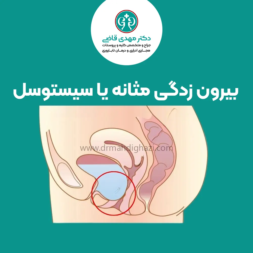
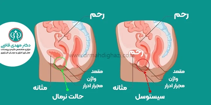
Under normal conditions, a woman’s bladder is held in place by strong pelvic muscles and tissues.
When these tissues become weak, the bladder can descend and bulge into the vagina; this is called bladder prolapse or cystocele.
In severe cases, the bladder drops so far that it bulges at the vaginal opening and becomes visible; this is called prolapse or protrusion.
Bladder prolapse is more common in older women.
Symptoms of bladder prolapse
The most common symptom is a bulge in the vagina that may be visible or felt.
Other symptoms and signs that may be related to bladder prolapse include:
Frequent urination or a feeling of urgency to urinate
Urinary incontinence
A feeling that the bladder does not empty completely
Recurrent urinary tract infections
Pain and heaviness in the vagina, pelvis, lower abdomen, groin, or lower back
Painful intercourse
In some cases, prolapse may cause no symptoms.
Causes of cystocele
Difficult and prolonged deliveries can put pressure on the bladder and cause bladder prolapse.
Other factors that can lead to pelvic prolapse include:
Lifting heavy objects
Chronic cough, or other lung problems
Constipation and repeated straining during bowel movements
Obesity
Menopause, when estrogen levels decrease
Estrogen helps strengthen the pelvic floor muscles.
When estrogen levels fall after menopause, these muscles become weaker and the risk of bladder prolapse increases.
Previous pelvic surgery: pelvic surgeries can weaken the muscles and support tissues of the bladder.
Aging: with age, pelvic floor muscles naturally become weaker, increasing the risk of bladder prolapse.
Diagnosis of bladder prolapse
Examination for bladder prolapse, or cystocele, is performed while the patient is lying down or standing, or while straining. The specialist assesses the degree of bladder prolapse and the possibility of other genital-area conditions.
In some women, in addition to bladder prolapse, the end of the large intestine or rectum may also prolapse and be visible or palpable; this condition is called rectocele.
Methods for diagnosing bladder prolapse
Different methods are used for accurate diagnosis of bladder prolapse, each providing useful information about the structure and function of the bladder and surrounding organs. The choice of diagnostic test depends on the patient’s symptoms and the severity of the condition.
Cystoscopy
In this method, an endoscope, a thin lighted camera, is inserted into the bladder under local anesthesia to evaluate the bladder wall.
Urodynamic testing
This test evaluates bladder function and urine flow.
X-ray
Sometimes the bladder is filled with contrast material through a catheter, and X-ray imaging is used to evaluate the bladder wall.
Ultrasound
Ultrasound gives the urologist a good image of the bladder, bladder volume, abnormalities of the inner bladder wall, and other organs inside the pelvis.
MRI imaging
In addition to bladder tissue, MRI clearly shows abnormalities of the pelvic floor muscles and other pelvic organs.
Treatment of bladder prolapse
Treatment of cystocele depends on the severity of symptoms and their effect on the patient’s life, and may include nonsurgical or surgical methods.
In mild cases, nonsurgical approaches such as observation without intervention, Kegel exercises, pelvic floor physical therapy, use of a pessary, and in some cases estrogen hormone therapy can help improve symptoms and prevent progression.
These methods are suitable options for patients with mild symptoms or for those who cannot undergo surgery because of age or health status.
In more severe cases, surgery may be needed to repair and strengthen the supportive tissues and correct bladder prolapse.
Several surgical methods are available for cystocele repair, including open surgery, minimally invasive surgery, and robot-assisted surgery. Depending on the type of surgery, biological or synthetic grafts or reinforcing materials may be used to strengthen the pelvic floor structure and improve prolapse symptoms.
Before deciding on surgery, the patient should be fully informed about the different aspects, risks, and benefits of this method. The urologic surgeon helps the patient make an informed decision by providing complete explanations and answering questions.
Nonsurgical treatment
Nonsurgical treatments for bladder prolapse are useful in mild cases and include the following:
Observation and no active treatment
Some women have bladder prolapse but no bothersome symptoms; therefore, they are followed every 6 months and treated if symptoms develop.
If the prolapse does not cause problems for the patient and does not block urine flow, treatment is not needed.
Behavioral treatment
This method may include the following:
Kegel exercises for bladder prolapse: These exercises are designed to strengthen the pelvic floor muscles around the vagina, bladder, and bowel.
Pelvic floor physical therapy: specialized physical therapy to strengthen the pelvic floor muscles
Use of a pessary: a vaginal pessary is a flexible device in different shapes that is placed inside the vagina to help prevent further prolapse and can be washed and replaced.
Medication treatment
Estrogen hormone therapy: reduced estrogen levels after menopause can weaken the pelvic floor muscles.
Surgical treatment
In more severe cases of bladder prolapse, surgery may be considered to repair and tighten the muscles and supportive tissues of the bladder.
The goal of surgery is to correct the prolapse, strengthen bladder-supporting tissues, and improve symptoms.
Surgery can be performed through the vagina or abdomen.
Surgical methods for bladder prolapse
Different surgical methods are available, including:
Open surgery: in this method, an incision is made in the lower abdomen or through the vagina, and the cystocele is repaired through that approach.
Minimally invasive surgery: laparoscopic instruments are used through 3 or 4 small openings in the lower abdomen to perform cystocele surgery.
Laparoscopic and robot-assisted surgery: in this method, robotic surgical instruments are inserted through the abdominal wall.
These instruments are connected to robotic arms and controlled by the surgeon.
For cystocele repair surgery, grafts or natural and synthetic tissues may sometimes be used:
Repair with a graft or natural tissue, in which a piece of the patient’s own tissue is taken from another part of the body and used for cystocele repair.
Reinforcement with synthetic surgical material or biological graft: when the patient’s own tissue is not sufficient, synthetic material or animal tissue may be used to strengthen the repair.
Before surgery, the patient should have a detailed discussion with the surgeon.
The patient should be informed about the risks, benefits, and other available options for surgical cystocele repair.
Obtaining informed consent from the patient is very important, and this consent is possible only after the urologist has answered all of the patient’s questions.
Course of bladder prolapse
Cystocele, or bladder prolapse, progresses gradually and may worsen over time, especially if risk factors such as pregnancy, repeated childbirth, and menopause are present. As the disease progresses, symptoms may change from mild to severe and may include heaviness and pain in the pelvic area, urinary problems such as frequent urination or involuntary urine leakage, and, in severe cases, difficulty emptying the bladder completely.
Cystocele can be divided into several stages:
In the early stages, prolapse is usually mild and may be asymptomatic or associated with very mild symptoms.
Over time, the prolapse reaches a moderate stage and symptoms become more obvious. The patient may feel more discomfort, especially when pressure is placed on the pelvic area, such as when lifting heavy objects or coughing.
In advanced stages, the prolapse can reach a point where part of the bladder is visible or can be felt through the vaginal opening. This stage usually causes severe discomfort and serious urinary problems and can significantly impair the patient’s quality of life.
Treatment outlook for cystocele
The treatment outlook for cystocele differs depending on the severity and stage of the disease and the body’s response to different treatments.
In mild and moderate stages, the outlook with nonsurgical methods such as pelvic floor exercises and pessary use is favorable and can often help reduce symptoms and prevent disease progression.
These treatments are recommended as initial options for patients with mild symptoms whose prolapse is not yet very severe.
In advanced cases, surgical treatment is usually needed to repair and strengthen pelvic floor tissues, control symptoms, and prevent further progression.
Surgeries often produce good results and can improve patients’ quality of life, but they may carry risks and sometimes repeat surgery is needed.
Overall, if patients monitor their condition regularly and use appropriate care and treatment methods, the long-term outlook for controlling cystocele and maintaining quality of life is favorable.
Summary
Cystocele, or bladder prolapse, is a type of prolapse in which the bladder bulges into the vagina because of weakness in the front wall of the vagina.
This condition, which occurs mostly in women, can result from repeated vaginal deliveries, older age, reduced estrogen after menopause, lifting heavy objects, and problems related to constipation or obesity.
Symptoms of cystocele include heaviness and pressure in the pelvic area, a bulge at the vaginal opening, urinary problems, and in more severe cases, urinary incontinence.
Diagnostic methods for bladder prolapse include pelvic examination, urine tests, and sometimes imaging tests to assess the severity of bladder prolapse.
Treatment of cystocele depends on the severity of the condition and can range from lifestyle changes and Kegel exercises to strengthen the pelvic floor muscles, to use of a pessary, a device that supports the bladder, and even surgery in more severe cases.
Regular pelvic floor exercises and attention to some care recommendations can help prevent disease progression and improve quality of life.
In this article, we tried to review the causes, symptoms, diagnostic methods, and treatment options for cystocele based on reliable scientific urology sources and explain effective ways to manage this condition.
Frequently asked questions
Actions & related links
Related articles
All articlesWhat Is Polycystic Kidney Disease? (Fetal and Adult PKD)
What is polycystic kidney disease? Learn about inherited ADPKD and ARPKD, fetal and adult symptoms, complications, diagnosis, medicines, surgery, diet, fluids, and prevention-focused care.
What Is a Renal Cortical Cyst? Symptoms, Diagnosis, and Treatment
Renal cortical cyst | Simple vs. complex kidney cysts | Warning symptoms | Diagnosis and treatment | Needle drainage, laparoscopy, medication, and ablation
Kidney Transplant: Cost, Blood Type Compatibility, and Surgical Method
What is kidney transplant? Learn about cost considerations, operation duration and method, diet, who may not be eligible, success rates, isolation precautions, and post-transplant care.
What Is Pyelonephritis? Kidney Infection Symptoms, Diagnosis, and Treatment
What is pyelonephritis? Learn kidney infection symptoms in women, children, men, and pregnancy; diagnosis, antibiotics, treatment, emergency warning signs, and cystitis differences.
Comments
0 comments
No comments yet. Be the first to share your thoughts.