
 Tap to zoom
Is Ultrasound or Cystoscopy Better for Diagnosing Bladder Cancer?
Is ultrasound or cystoscopy better for bladder cancer diagnosis? Learn the advantages, limitations, cystoscopy steps, and complementary urine and blood tests.
- Published on
- June 26, 2026
- Reading time
- 5 min read
- Last updated
- Updated: June 27, 2026
Ultrasound is a noninvasive, safe, and relatively low-cost method that can reveal early signs of bladder cancer, but it also has limitations. When ultrasound results are unclear or the physician suspects a lesion, cystoscopy is used. Cystoscopy is more precise than ultrasound and allows direct visualization of the inside of the bladder.
In this article, we aim to explain in simple, scientific terms how bladder cancer diagnosis with ultrasound is performed. We will also explain when bladder cystoscopy or other imaging methods may be needed. For accurate information about the diagnostic pathway for bladder cancer, we suggest reading this article to the end.
Urologist Bladder disease specialist
Note: To improve care quality and patient satisfaction, appointments are triaged by reason for visit. Each physician sees patients within the relevant urology subspecialty.
Book appointment
Why is accurate diagnosis of bladder cancer important?
Bladder cancer often appears with concerning symptoms such as blood in the urine (hematuria), unusual urinary frequency, or pain and burning during urination. Noticing these signs is the first step toward seeing a urologist and starting the diagnostic process.
The earlier the disease is found, the more effective and successful treatment measures can be. As a result, the patient's prognosis improves substantially. Delay in diagnosis can allow the cancer to progress into deeper layers of the bladder or spread to other parts of the body, making treatment much more difficult and complex.
Physicians use several different methods to diagnose bladder cancer. These methods include:
Appointments related to Urologist Bladder disease specialist
Open the public booking path to review times and related information.
Book appointment
Urine and blood tests (for screening and assessment of overall health);
Ultrasound (as an initial noninvasive imaging method);
Cystoscopy (as the gold standard for bladder cancer diagnosis, with the ability to take a biopsy);
Each of these methods has its own advantages and limitations, and the choice depends on the patient's clinical condition.
Bladder cancer diagnosis with ultrasound

Bladder cancer diagnosis with ultrasound is one of the first and most common steps in evaluating patients suspected of having a bladder tumor. In this method, the radiologist uses sound waves, without surgery or inserting instruments into the body, to observe the shape and condition of the bladder.
During bladder cancer diagnosis with ultrasound, the radiologist or sonographer moves the ultrasound probe over the lower abdomen. This allows the physician to see the bladder wall and any abnormal mass or protrusion, such as a tumor, on the monitor. The advantages of ultrasound are described below:
Q&A — Urologist Bladder disease specialist
General questions are shown on the destination page after review.
Book appointment
Noninvasive and painless: performed without a surgical incision or placing an instrument inside the body.
No ionizing radiation: unlike CT scan or X-ray, ultrasound does not use radiation.
Accessible and cost-effective: it is usually easy to perform in most diagnostic centers and has a relatively reasonable cost.
Ultrasound also has limitations, which are noted below:
Dependent on the radiologist's skill: image quality and interpretation depend on the radiologist's skill and experience.
Failure to detect small masses: very small tumors or flat superficial lesions in the bladder wall may not be shown well.
Inability to assess depth of invasion: it cannot definitively determine how deeply a tumor has penetrated the different layers of the bladder wall.
Accuracy of ultrasound in diagnosing bladder cancer in early stages
In the early stages of bladder cancer diagnosis, ultrasound is accurate enough to identify many sizable masses; however, errors are possible with tumors smaller than 5 millimeters or with superficial lesions.
Given these limitations, ultrasound is often used as an initial screening tool or an adjunct method to evaluate lesions seen by other techniques. If a patient has blood in the urine but ultrasound is negative, the urologist will recommend another complementary method, such as cystoscopy, for confirmation.
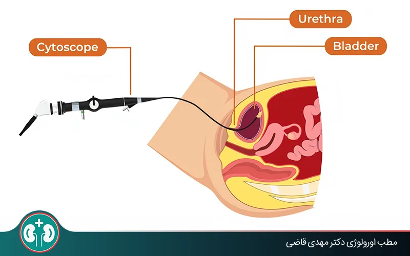
Bladder cystoscopy: an accurate method for examining the inside of the bladder

Bladder cystoscopy is considered the best method for diagnosing bladder cancer. In this method, the urologist uses a thin, flexible instrument equipped with a camera and light, called a cystoscope, to look directly inside the bladder. This process is usually performed in an outpatient setting with local anesthesia.
Cystoscopy allows the urologist to see the bladder wall directly and with high magnification. If needed, tissue from suspicious areas can be sampled for bladder cancer pathology. When ultrasound or other methods are inconclusive, cystoscopy helps make an accurate diagnosis and reduces the chance of error.
Steps of cystoscopy
Cystoscopy for bladder cancer is a precise, targeted process that is usually performed in the following order:
1. Preparation:First, the patient lies on the examination bed. To reduce discomfort, the urologist injects a local anesthetic gel into the urethra about 30 minutes before cystoscopy. Before inserting the cystoscope, other gels are also usually applied so the cystoscopy proceeds more smoothly and anesthesia is enhanced.
2. Inserting the cystoscope:The urologist gently inserts the cystoscope through the urethra into the bladder. Cystoscopy can also be performed under anesthesia, especially if the physician intends to treat or take a sample in the hospital.
3. Internal examination of the bladder:The physician fills the bladder with fluid, usually sterile water or normal saline, so its walls open fully and the inner lining can be seen clearly. The inner walls are then examined for any mass, ulcer, tumor, or abnormal area.
4. Biopsy, if needed:If a suspicious lesion is seen during cystoscopy, the urologist can take a biopsy using very small instruments passed through the cystoscope. The sample is sent to the laboratory for more precise evaluation.
5. End of the procedure:After complete examination, the fluid is drained and the cystoscope is removed. The entire cystoscopy procedure usually takes 10 to 45 minutes.
Advantages and limitations of cystoscopy
Overall, bladder cystoscopy is slightly more invasive than ultrasound, but it plays a key role in definitive diagnosis of tumors and determining their type. This method is usually performed after imaging studies so a final and reliable result can be obtained. The advantages and limitations of cystoscopy are listed below.
Advantages of cystoscopy:
High diagnostic accuracy;
Direct visualization of the inside of the bladder;
Assessment of tumor location and appearance;
Ability to take a biopsy;
Help with early diagnosis and effective treatment.
Disadvantages of cystoscopy:
Minimally invasive nature: it requires insertion of the cystoscope into the body.
Risk of infection: there is a possibility of infection, and for this reason antibiotics are prescribed for everyone who undergoes cystoscopy.
Temporary side effects: it may cause discomfort, burning with urination, or a small amount of blood in the urine for one or two days.
Higher cost than other bladder cancer diagnostic methods: cystoscopy requires specialized instruments and must be performed by a urologist; therefore it costs more than other bladder cancer diagnostic methods.
Difference between ultrasound and cystoscopy in bladder cancer diagnosis

The main difference between ultrasound and cystoscopy is in how they are performed, their accuracy, and their role in bladder cancer diagnosis. Ultrasound is a noninvasive and inexpensive method for initial screening, whereas cystoscopy is a minimally invasive and highly accurate method for direct visualization inside the bladder and biopsy. The table below fully compares these two bladder cancer diagnostic methods:
Feature | Bladder ultrasound | Cystoscopy |
|---|---|---|
How it is performed | Noninvasive: performed from the abdomen using sound waves and an ultrasound machine | Minimally invasive: the physician inserts a thin tube called a cystoscope through the urethra into the bladder |
Accuracy | Relatively lower accuracy; it depends on the technician's skill and tumor size. | Very accurate and the gold-standard method for diagnosing bladder cancer |
Cost | A cost-effective method for initial screening | It has a higher cost because it requires specialized equipment and an operating-room setting. |
Use | Initial screening and evaluation of masses or kidney and bladder problems (the first diagnostic step) | Definitive diagnosis, determination of tumor size and location, and biopsy for pathology tests |
Biopsy | Not possible | Allows sampling of suspicious tissue for evaluation of the lesion |
Additional notes | Painless and does not require special preparation | Performed with local anesthesia, with possible burning and mild bleeding afterward |
Complementary tests for bladder cancer diagnosis (urine and blood)
In the early evaluation of symptoms suspicious for bladder cancer, urine and blood tests can provide clues about disease or the person's overall health; however, final diagnosis always requires specialized imaging methods, such as ultrasound, and ultimately cystoscopy and biopsy. Their role in bladder cancer diagnosis is reviewed below.
Bladder cancer diagnosis in urine testing
One of the most common symptoms of bladder cancer is blood in the urine. This blood may be visible or detected only under a microscope during urinalysis. Urine cytology is a specialized test in which a urine sample is examined under a microscope to identify abnormal or cancerous cells shed from the bladder wall.
Urine cytology is very useful for diagnosing high-grade tumors because these tumors are more likely to shed cells into the urine. This test is less accurate for low-grade tumors, and a negative cytology result alone does not rule out cancer.
Blood test for bladder cancer diagnosis

Blood testing is not used to diagnose bladder cancer, but it plays an important role in monitoring, determining prognosis, and assessing the patient's overall health. The following blood-test factors are considered by the urologist during bladder cancer diagnosis:
Complete blood count (CBC):This factor is used to assess anemia, which may result from chronic bladder bleeding, and to evaluate platelets and white blood cells.
Blood chemistry tests: these tests evaluate kidney and liver function. Bladder cancer can obstruct the urinary tract, so assessment of kidney function, including creatinine level, is especially important.
In addition, blood tests can be used to measure levels of specific markers, such as certain proteins or inflammatory markers. These tests and evaluations are useful during chemotherapy or other treatments for monitoring the body's response to treatment and side effects.
Screening and stages of bladder cancer diagnosis
Bladder cancer diagnosis usually follows the appearance of early symptoms and proceeds through several stages. There is no general screening program for bladder cancer. However, people at high risk should pay close attention to their symptoms. Tobacco users and people exposed to industrial dyes are considered risk groups. The diagnostic process is often as follows:
1. Appearance of symptoms: the first step is usually noticing symptoms such as blood in the urine (hematuria), burning or pain during urination, and urinary frequency.
2. Ultrasound or CT scan: after symptoms appear, the urologist may order ultrasound or CT scan to evaluate the kidneys, bladder, and other pelvic organs. These methods help identify large, prominent masses in the bladder.
3. Complementary tests: at this stage, urine tests, especially urine cytology, and blood tests are performed to look for abnormal cells and assess overall health.
4. Cystoscopy: if the results of previous steps raise suspicion for bladder cancer, cystoscopy is performed as the gold-standard method. In this method, the urologist directly views the inside of the bladder, examines any mass or abnormality, and takes a biopsy for definitive diagnosis. Biopsy is the only way to definitively confirm the presence of cancer cells.
Prognosis after bladder cancer diagnosis

Bladder cancer prognosis depends on the stage and depth of tumor involvement. In early stages, when the tumor has not yet invaded the bladder muscle, the chance of successful treatment is very high. In more advanced stages, combined treatments such as surgery, chemotherapy, or immunotherapy are needed. Early diagnosis with ultrasound and cystoscopy can play a vital role in improving prognosis and increasing patient survival.
Cost of bladder cystoscopy
Despite its high accuracy and importance, cystoscopy, like any other medical procedure, has its own costs and side effects that the patient should know about. The cost of bladder cystoscopy depends on several factors, including the type of cystoscopy, the type of hospital or clinic (public, private, or specialized), insurance coverage, and physician fees.
The type of cystoscopy refers to whether the procedure is performed with local anesthesia, sedation, or general anesthesia, each of which has a different cost. In addition, taking a biopsy increases the total cost.
Summary
Ultrasound is one of the first and simplest methods for evaluating the bladder and initially detecting suspicious masses. However, cystoscopy and complementary urine and blood tests are necessary to confirm the diagnosis and determine the type of lesion. Combining these methods helps the urologist identify bladder cancer in early stages and choose the best treatment decision.
Diagnosing bladder cancer in early stages greatly increases the chance of successful treatment. Therefore, if people experience symptoms such as blood in the urine, unusual urinary frequency, or burning during urination, seeing a urologist and completing the necessary evaluations is the best way to protect their health.
Frequently Asked Questions
Actions & related links
Related articles
All articlesWhat Is Polycystic Kidney Disease? (Fetal and Adult PKD)
What is polycystic kidney disease? Learn about inherited ADPKD and ARPKD, fetal and adult symptoms, complications, diagnosis, medicines, surgery, diet, fluids, and prevention-focused care.
What Is a Renal Cortical Cyst? Symptoms, Diagnosis, and Treatment
Renal cortical cyst | Simple vs. complex kidney cysts | Warning symptoms | Diagnosis and treatment | Needle drainage, laparoscopy, medication, and ablation
Kidney Transplant: Cost, Blood Type Compatibility, and Surgical Method
What is kidney transplant? Learn about cost considerations, operation duration and method, diet, who may not be eligible, success rates, isolation precautions, and post-transplant care.
What Is Pyelonephritis? Kidney Infection Symptoms, Diagnosis, and Treatment
What is pyelonephritis? Learn kidney infection symptoms in women, children, men, and pregnancy; diagnosis, antibiotics, treatment, emergency warning signs, and cystitis differences.
Comments
0 comments
No comments yet. Be the first to share your thoughts.